برای تماس کلیک کنید

سما تشخیص آریا ، عرضه کننده متنوع ترین و کامل ترین محصولات کیت های آزمایشگاهی

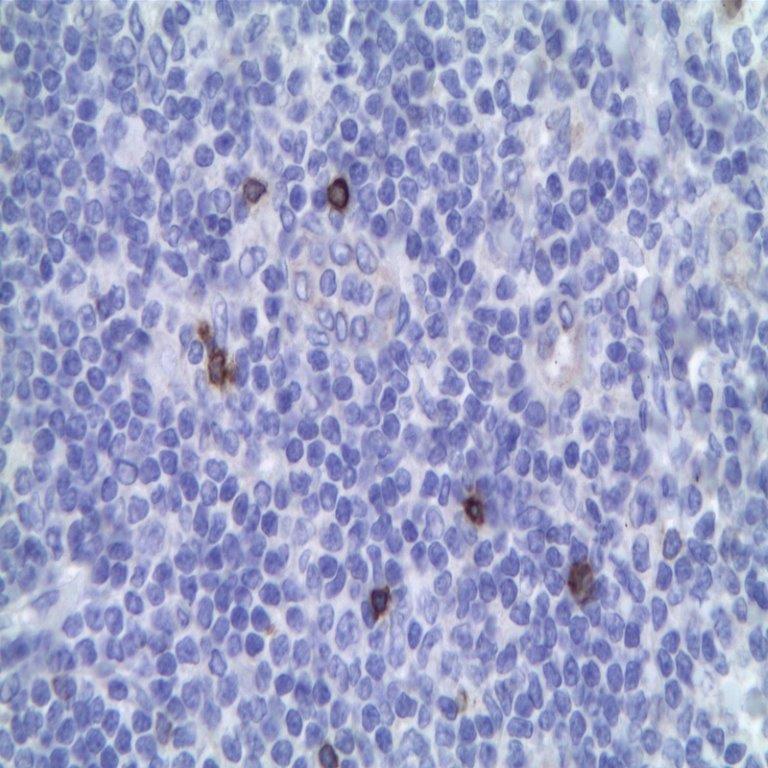
آنتی­ بادی TCR C Gamma M1 (H-41)

Name: TCR gamma M1 Antibody Clone H-41

Description and applications: The antibody detects TCR C gamma M1 which has a predicted molecular
weight of approximately 18 kDa.
The ability of T cell receptors (TCR) to discriminate foreign from self-peptides presented by major histocompatibility complex (MHC) class II molecules is essential for an effective adaptive immune response. TCR recognition of self-peptides has been linked to autoimmune disease. Mutant self-peptides have been associated with tumors. Engagement of TCRs by a family of bacterial toxins know as superantigens has been responsible for toxic shock syndrome. Autoantibodies to V beta segments of T cell receptors have been isolated from patients with rheumatoid arthritis (RA) and systemic lupus erythematosus (SLE). The autoantibodies block TH1-mediated inflammatory autodestructive reactions and are believed to be amethod by which the immune system compensates for disease.
T Cell and TCR Diversity Most human T cells express the TCR alpha-beta and either CD4 or CD8 molecule (single positive, SP). A small number of T cells lack both CD4 and CD8 (double negative, DN). Increased percentages of alpha-beta DN T cells have been identified in some autoimmune and immunodeficiency disorders. Gamma-delta T cells are primarily found within the epithelium. They show less TCR diversity and recognize antigens differently than alpha-beta T cells. Subsets of gamma-delta T cells have shown antitumor and immunoregulatory activity.
The antibody has a particular utility in the diagnostic of the four major subtypes of predominantly γδ T-cell lymphomas that are currently recognized: 1) hepatosplenic γδ T lymphoma; 2) Subcutaneous panniculitis-like T cell lymphoma; 3) primary cutaneous γδ T cell lymphoma and 4) IInd subtype of lymphoma associated with enteropathy. However, the antibody against the gamma chain of the TCR molecule can be positive in isolated case of mycosis fungoides or lymphomatoid papulosis. Consequently, the clinical and histological context in which the lesion occurs together with other immunocytochemical markers, must be taken into account when specifying the diagnosis.

Composition: anti-human TCRgamma mouse monoclonal antibody purified from serum and prepared in 10mM PBS, pH 7.4, with 0.2% BSA and 0.09% sodium azide

 Immunogen: Human TCR gamma chain constant region.

حجم موجود3 / 7 / 12 میلی‌لیتر

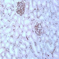
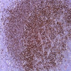
مورد استفادهایمونوهیستوشیمی روی بافت پارافین. بر روی بافتهای فروزن یا وسترن بلات تست نشده است.

کلونH-41

ایزوتایپ: IgG1K

کنترل مثبت IHC: تونسیل

قابلیت دیده شدن: سیتوپلاسمی

روش پیشنهادی IHC:

  • برش تهیه شده با ضخامت μm4 باید روی لام شارژ شده در طول شب و در دمای 60 درجه سانتی گراد خشک شود.
  • دپارافینه کردن، رهیدراته کردن و استفاده از حرارت جهت بازیابی اپی توپ : بافت را با استفاده از بافر EDTA pH 8 برای بیست دقیقه در دمای 95 درجه سانتی گراد بجوشانید. سپس 5-3 بار با استفاده از آب مقطر یا دیونیزه شده شسته و سپس بیست دقیقه در دمای اتاق خنک کنید.
  • بلاک پروکسیدازهای درونی : بلاک با استفاده از محلول پروکسیداز به مدت ده دقیقه
  • آنتی بادی اولیه: برای 10 دقیقه در انکوبه کنید پروتکل ممکن است بسته به تهیه نمونه و کاربرد خاص متفاوت باشد. شرایط استاندارد باتوجه به آزمایشگاه و کاربر متفاوت می باشد.
  • برای رنگ آمیزی ، می توانید از کیت Master Polymer Plus Detection یا کیتهای دیتکشن سایر کمپانی ها استفاده کنید.
  • رنگ میزی نهایی با هماتوکسیلین و فیکس نهایی اسلاید.

شرایط نگهداری و پایداری : حداکثر 18 ماه؛ در دمای 2-8 درجه سانتیگراد نگهداری شود. فریز نشود.


Download Cataloge

Icon

TCR C Gamma M1 (H-41) 349.19 KB 8 downloads

...
.well.c2a3 .btn { padding: 11px 30px; font-size: 11pt; } .well.c2a3 .media-body { font-size: 11pt; } .well.c2a3 .wpdm_icon { height: 42px; width: auto; }

Reviews

There are no reviews yet.

Be the first to review “آنتی­ بادی TCR C Gamma M1 (H-41)”

Your email address will not be published. Required fields are marked *

Scroll to Top