-
 آنتی بادیهای ایمونوهیستوشیمی
آنتی بادی Cytokeratin 7 (OVTL 12/30)
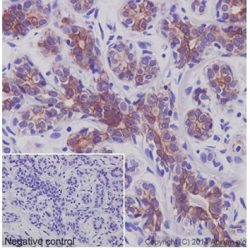
Name: Cytokeratin 7 Antibody clone OV-TL 12/30
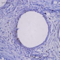
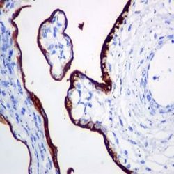
Description and aplications: Keratins (also known as cytokeratins) are intermediate filament/forming proteins that represents the main cytoskeleton of epithelial cells. They classically are classified based on Moll cataloging which grouped the basic-to-neutral type II keratins as K1–K8 and the acidic type I keratins as K9– K19. An updated classification including 24 types of keratins was then proposed in order to enable other mammalian species keratins to be added. This monoclonal antibody reacts specifically with the cytokeratin 7, immunostaining of the producing a protein band of 54 kD in the cytoskeleton of cell lines and uni-dimensional immunoblots. This antibody is useful to distinguish between different types of normal glandular epithelia as it marks the epithelia of lung and breast, being negative for colon and prostate. We have not observed any cross reaction with other cytokeratins and overall this antibody does not react with stratified squamous epithelium. In liver, hepatocytes are negative and the epithelial cells of the bile ducts are positive. This antibody reacts with numerous both benign and malignant epithelial lesions. Cytokeratin 7 is expressed in specific subtypes of ovarian adenocarcinomas, breast and lung carcinomas while the gastrointestinal tract, except stomach, are negative. Carcinomas arising from transitional epithelium also express cytokeratin, whereas prostate cancer is generally negative.
Composition: anti human keratin 7 mouse monoclonal antibody obtained from supernatant culture and prediluted in a Tris buffered solution pH 7.4 containing 0.375mM sodium azide solution as bacteriostatic and bactericidal.
Intended use : Immunohistochemistry (IHC) on paraffin embedded tissues. Not tested on frozen tissues or Western-Blotting
Immunogen: OTN II ovarian carcinoma cell line
-
 آنتی بادیهای ایمونوهیستوشیمی
آنتی بادی Cytokeratin 8 (EP17)
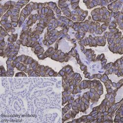
Name: Cytokeratin 8 antibody clone EP17
Description and aplications: Cytokeratin 8 (CK8) is an intermediate filament protein produced early in embryogenesis. It is the only type-II CK occurring in many simple epithelial in respiratory, gastrointestinal, male and female reproductive tract and thyroid. CK8 is often co-expressed with Cytokeratin 18. CK8/18 is the major keratin pair in simple-type epithelia, as found in the liver, pancreas, and intestine. CK8 antibody is used to detect adenocarcinomas with simple epithelium origin. The difference in staining pattern is useful to distinguish duct (peripheral staining) from lobular (perinuclear staining) breast carcinoma
Composition: anti-human cytokeratin 8 rabbit monoclonal antibody purified from ascites. Prepared in 10mM PBS, pH 7.4, with 0.2% BSA and 0.09% sodium azide
Immunogen: A synthetic peptide corresponding on the C-terminus
-
 آنتی بادیهای ایمونوهیستوشیمی
آنتی بادی Cytokeratin 8/18 (B22.1&B23.1)
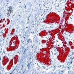
Name : Cytokeratin 8/18 antibody clone B22.1/B23.1
Description and applications:Cytokeratins 8&18 can be found in most simple epithelium,e.g. thyroid,female breast, gastrointestinal tract, and respiratory tract. Adenocarcinomas and most non-keratinizing squamous carcinomas will stain, but keratinizing squamous carcinomas will not. This antibody is used when attempting to demonstrate the presence of Paget cells; there is very little keratin 18 in the normal epidermis so this will only stain Paget cells. This approach facilitates the interpretation using immunostains and is more sensitive than mucin histochemistry.
Composition:Anti-human Cytokeratin 8/18 cocktail of two mouse monoclonal antibodies purified from serum and prepared in 10mM PBS, pH 7.4, with 0.2% BSA and 0.09% sodium azide
Intended use : Immunohistochemistry (IHC) on paraffin embedded tissues. Not tested on frozen tissues or Western-Blotting
-
 آنتی بادیهای ایمونوهیستوشیمی
آنتی بادی Cytokeratin 8/18/19 (B22.1&B23.1/BA17)
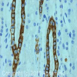
Name: Cytokeratin 8/18/19 Antibody clone B22.1&B23.1/BA17
Description and application: Keratins are cytoplasmic intermediate filament proteins expressed by epithelial cells. Cytokeratins 8 & 18 (CK 8 & 18) can be found in most simple epithelia, e.g. thyroid, female breast, gastrointestinal tract, and respiratory tract. Adenocarcinomas and most non-keratinizing squamous carcinomas will stain with anti-CK 8 & 18, but keratinizing squamous carcinomas will not. This antibody is used when attempting to demonstrate the presence of Paget cells; there is very little keratin 18 in the normal epidermis so this will only stain Paget cells. The use of immunostaining facilitates the interpretation and has been shown to be more sensitive than mucin histochemistry. Cytokeratin 19 is a member of type I acidic subfamily of intermediate filaments. It is expressed in various different human tissues except in liver. Keratin 19 is not expressed in hepatocytes, therefore, antibody to keratin 19 is useful in the identification of liver metastasis.
Composition: Anti-human Cytokeratin 8/18/19 Mouse monoclonal antibody prepared in 10mM PBS, pH 7.4, with 0.2% BSA and 0.09% sodium azide.
Intended use: Immunohistochemistry (IHC) on paraffin embedded tissues. Not tested on frozen tissues or Western-Blotting
-
 آنتی بادیهای ایمونوهیستوشیمی
آنتی بادی Myeloperoxidase (Polyclonal)
Name: Myeloperoxidase antibody
Description and application: Myeloperoxidase is an important enzyme used by granulocytes during phagocytic lysis of foreign particles engulfed. In normal tissues and in a variety of myeloproliferative disorders myeloid cells of both neutrophilic and eosinophilic types, at all stages of maturation, exhibit strong cytoplasmic reactivity for MPO. Erythroid precursors, megakaryocytes, lymphoid cells, mast cells, and plasma cells are nonreactive. MPO is not observed in the neoplastic cells of a wide variety of epithelial tumors and sarcomas. MPO is useful in differentiating between myeloid and lymphoid leukemias.
Composition: Anti-human Myeloperoxidase rabbit polyclonal antibody purified from serum and prepared in 10mM PBS, pH 7.4, with 0.2% BSA and 0.09% sodium azide
Intended use: Immunohistochemistry (IHC) on paraffin embedded tissues. Not tested on frozen tissues or Western-Blotting
-
 آنتی بادیهای ایمونوهیستوشیمی
آنتیبادی Placental Alkaline Phosphatase (SP15)
Name: Placental Alkaline Phosphatase antibody
Descrioption and aplication: This antibody reacts with a membrane-bound isoenzyme of placental alkaline phosphatase (PLAP) occurring in the placenta during the 3rd trimester of gestation. It is expressed in testicular germ cell tumors. Unlike germ cell tumors, PLAP-positive somatic cell tumors uniformly express epithelial membrane antigen (EMA).
Composition: Anti-human Placental Alkaline Phosphatase rabbit monoclonal antibody purified from serum and prepared in 10mM PBS, pH 7.4, with 0.2% BSA and 0.09% sodium azide
Intended use : Immunohistochemistry (IHC) on paraffin embedded tissues. Not tested on frozen tissues or Western-Blotting
Immunogeon: Recombinant protein encoding human placental alkaline phosphatase.
-
 آنتی بادیهای ایمونوهیستوشیمی
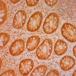
آنتی بادی SATB2 (DNA-Binding Protein SATB2) (EP281)
Name: DNA-Binding Protein SATB2 Monoclonal Antibody clone EP281
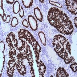
Description and applications: DNA-binding protein SATB2, also known as Special ATrich sequence-binding protein 2, is a nuclear matrixassociated transcription factor. SATB2 acts as a docking site for chromatin remodelling enzymes and recruits co-activators and co-repressors to control nuclear gene expression. SATB2 also regulates skeletal development, osteoblast differentiation, and modulates immunoglobulin expression. In normal tissues, strong nuclear SATB2 expression is observed in essentially all glandular cells lining the lower gastrointestinal tract, including the appendix, colon, and rectum. SATB2 is also expressed in a subset of neuronal cells from the cerebral cortex and hippocampus. In tumor tissues, SATB2 is detected in cancer cells of colorectal origin and may function as a clinically useful diagnostic marker for colorectal cancer (CRC). In a multi-cohort study with 1882 primary and metastatic CRCs, SATB2 shows high sensitivity (85%) for CRC, and further enhanced to 93% when stained in conjunction with Cytokeratin 20. A recent study showed SATB2 expression in 89% of medullary carcinomas of the large intestine. SATB2 has been suggested as a valuable prognostic marker: high SATB2 expression was determined as an independent marker of good prognosis and sensitivity to chemotherapy and radiation in CRC while loss of SATB2 expression was correlated with poor prognosis in laryngeal carcinoma patients.
Composition: Anti-human SATB2 rabbit monoclonal antibody purified from serum and prepared in 10mM PBS, pH 7.4, with 0.2% BSA and 0.09% sodium azide
Intended use: Immunohistochemistry (IHC) on paraffin embedded tissues. Not tested on frozen tissues or Western-Blotting
-
 آنتی بادیهای ایمونوهیستوشیمی
آنتی بادی Carcinoembryonic Antigen (CEAm) (COL-1)
Name: CEAm Antibody clone COL-1
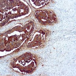
Description and applications: This antibody has a high affinity for CEA and shows no detectable reactivity to nonspecific cross-reacting antigen (NCA), biliary glycoprotein (BGP) and human polymorphonuclear leucocytes. Ab-3 shows no reaction with a variety of normal tissues .CEA is not found in benign glands, stroma, or malignant prostatic cells. Antibody to CEA is useful in detecting early foci of gastric carcinoma and in distinguishing pulmonary adenocarcinomas (60-70% are CEA+) from pleural mesotheliomas (rarely or weakly CEA+).
Composition: anti-human CEA mouse monoclonal antibody purified from ascites. Prepared in 10mM PBS, pH 7.4, with 0.2% BSA and 0.09% sodium azide
Intended use: Immunohistochemistry (IHC) on paraffin embedded tissues. Not tested on frozen tissues or Western-Blotting
-
 آنتی بادیهای ایمونوهیستوشیمی
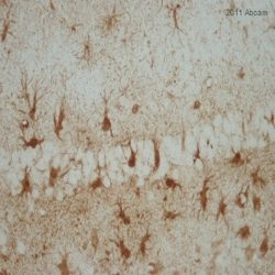
آنتی بادی S100-A1 Protein (EP184)
Name: S100-A1 Protein Antibody clone EP184
Description and applications: The S100-A1 protein belongs to the S100 protein family, a set of 25 small acidic molecules with a molecular mass of 10-12 kDa that are functionally grouped into homo or heterodimers composed of two subunits, alpha and beta, with extensive sequence homology and capable of combining with each other. There exist up to 14 variants of the alpha chain gene, which is located on chromosome 1q21; on the contrary, there is only one sequence of the beta chain gene, located on chromosome 21q22.3, showing its greater functional specificity. Depending on the combination between the alpha and beta single chains and the homology of their sequences, there exist three forms of S-100 protein: A, with at least 15 subtypes between A1 and A15, B, and G. In their dimeric form, S100 proteins, together with calmodulin and troponin C, belong to the calcium binding protein family; their affinity for this ion, as well as for other metals such as zinc, is remarkable. It is for this reason that the S100 protein is involved in numerous basic cellular activities such as cation diffusion across lipid membranes, microtubule assembly, and regulation of RNA polymerase activity. In neurons, the S100 protein also regulates the interaction between chromosomes and synaptosomes. As for the S100-A1 protein, its most important role is to regulate myocardial contractility. In normal tissues, S100-A1 protein expression is observed at the sarcolemma of the cardiac and the skeletal muscle, in a smaller proportion in the latter; likewise, this protein is found in renal tubule and brain cells. It is not expressed in other tissues. In neoplasms, S100-A1 protein expression has been investigated predominantly in renal tumour pathology, where it is important for the diagnosis of oncocytic tumours, both oncocytomas and oncocytic papillary carcinomas, and allows the differential diagnosis with chromophobe carcinomas, especially its eosinophilic variant, which does not express this protein. For this reason, the S100-A1 protein, together with cytokeratin 7, vimentin, and c-kit (CD117) has been included in the optimal panel for the diagnosis of renal oncocytomas. Additionally, and compared to normal urothelium, which may exhibit very slight staining, nephrogenic adenomas show intense cytoplasmic and/or nuclear/cytoplasmic staining. For all of the above reasons, the S100-A1 protein, together with PAX8, p63, PSA, and CEA, is a highly sensitive and specific marker for the diagnosis of nephrogenic adenomas, making it a useful marker in the differential diagnosis of male urinary and genital system tumours, including prostatic adenocarcinomas, which are regularly negative for this protein. The S100-A1 protein is also useful in the differential diagnosis between dysplastic melanocytic nevi and malignant melanoma, with slight staining in the former and more intense staining in the latter, in contrast to the S100-B protein, which tends to
present greater expression in nevus lesions, except in areas of melanoma with high proliferation. Finally, in both endometrial and ovarian endometrioid carcinomas, S100-A1 protein overexpression is an
indicator of a poorer prognosi .Composition: Anti-human S100-A1 Protein rabbit monoclonal antibody purified from serum and prepared in 10mM PBS, pH 7.4, with 0.2% BSA and 0.09% sodium azide
Intended use: Immunohistochemistry (IHC) on paraffin embedded tissues. Not tested on frozen tissues or Western-Blotting
Clone: EP184
Ig isotype: IgG
IHC positive control: Myocardium, skeletal muscle, or renal cell carcinoma tissue section.
Visualization: Cytoplasm.
-
 آنتی بادیهای ایمونوهیستوشیمی
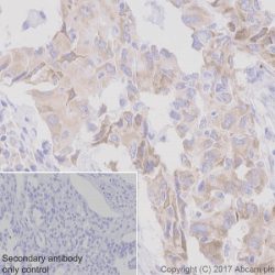
آنتی بادی ROS1 (Proto-Oncogene Tyrosine-Protein Kinase ROS)(D4D6)
Name: Proto-Oncogene Tyrosine-Protein Kinase Antibody clone D4D6
Description and applications: The transmembrane proto-oncogene tyrosine- kinase protein ROS, better known as ROS1, is a protein belonging to the insulin receptors tyrosine-kinase subfamily whose production is encoded by the ROS (MCF3) gene, located in the 6q22.1 chromosome region. After its activation, this gene intervenes in numerous molecular pathways related to the cell differentiation, proliferation, growth and survival including the activation of the PI3K-mTOR pathway. Chromosome aberrations affecting the ROS gene have been described in the glioblastoma multiforme through fusions of the C-terminal portion of ROS1 with the N-terminal domain of the FIG (Fused in Glioblastoma) encoded in the GOPC gene. The chimeric protein GOPC-ROS1 is located at the level of the Golgi apparatus and presents activity as tyrosine kinase receptor. Other fusions of ROS1 with different genes such as SLC34A2, CD74, EZR, LRIG3, SDC4, TPM3, CCDC6 or KDELR2 among others, have as a result the appearance and expression of several chimeric proteins in 1-3 % of cases of lung adenocarcinoma and represents the therapeutic target for Crizotinib and analog molecules. Non-specific staining on macrophages and reactive type II pneumocytes has also been described in isolated cases, whereas mucinous adenocarcinomas in general show low diffuse cytoplasmic staining in the absence of the translocation of the ROS1 gene. In order to identify the cases of lung carcinomas with translocations of ROS1, complex techniques of RT-PCR and FISH can be used, although the rabbit monoclonal antibody D4D6 has been recently validated as an useful screening tool for positive cases of immunostaining against ROS1, which in comparative studies through in situ hybridization techniques with break-apart probes has proven a sensitivity and specificity of over 95%. In order to consider a case as positive for ROS1, the immunostaining has to be strong or moderately intense on the membrane and/or the cytoplasm in more than 75% of tumor cells. Depending on the type of fusion of the ROS1 gene, other immunostaining patterns can be expressed as cytoplasmic punctiform in the CD74-ROS1 cases or cytoplasmic with linear accentuation in the lateral or apical membrane in the EZR-ROS1 cases. Although the morphology of the tumor cannot be considered as a criterion to select the positive ROS1 cases, the solid, micro-papillary, cribriform and signet ring cell growth patterns have been observed more frequently among the positive ROS1 cases. Focal and low-intensity staining can also be observed in up to 30% of the non-translocated cases. Therefore, the result must be confirmed with other analytical methods. 50% of the inflammatory myofibroblastic tumors also present translocation of the ALK gene, whereas the remaining ones show a little known genetic profile. It has been recently proven in a relatively low number of cases that up to 10% of these tumors show diffuse cytoplasmic or punctiform staining against the D4D6 clone (with translocation confirmed through FISH and RT-PCR techniques). In this same study, the antibody proved weak, focak nuclear staining in cases of gastrointestinal stromal tumors, myofibroblastic sarcomas, leiomyosarcomas and follicular dendritic cell sarcomas.Less than 1% of colorectal adenocarcinomas and up to 5% of stomach adenocarcinomas can show translocations of the ROS1 gene and consequential cytoplasmic staining. The low percentage of colorectal adenocarcinomas makes the gene as a potential therapeutic target, but in the case of the stomach adenocarcinomas, it can represent a therapeutic alternative, since, in general, the positive ROS1 cases show a non-amplified phenotype of HER2 and MET.
Composition: Anti-human ROS1 rabbit monoclonal antibody purified from serum and prepared in 10mM PBS, pH 7.4, with 0.2% BSA and 0.09% sodium azide
Immunogen: Human colon carcinoma extract.
Species reactivity: In vitro diagnostics in humans. Not tested in other species
-
 آنتی بادیهای ایمونوهیستوشیمی
آنتی بادی SDHB (EP288)
Name: SDHB Monoclonal Antibody clone EP288
Description and applications: Succinate dehydrogenase (SDH) is Complex II in the mitochondria, vital for mitochondrial electron transport, as well as Krebs cycle function. SDH catalyzes the oxidation of succinate to fumarate and transfers electrons to ubiquinone through the coordination of its four subunits (SDHA, SDHB, SDHC, and SDHD). The SDH complex functions as a tumor suppressor. Loss of any subunit proteins lead to destabilization of the complex and tumor formation. SDH subunit B (SDHB) is ubiquitously expressed in normal tissues. Germline mutations in SDHB, SDHC, or SDHD genes predisposes development of phaeochromocytoma, paraganglioma and gastrointestinal stromal tumor (GIST). SDHB immunohistochemistry is helpful in identification of phaeochromocytomas, paragangliomas or GIST with SDHB mutation.
Composition: Anti-human SDHB rabbit monoclonal antibody purified from serum and prepared in 10mM PBS, pH 7.4, with 0.2% BSA and 0.09% sodium azide
Intended use: Immunohistochemistry (IHC) on paraffin embedded tissues. Not tested on frozen tissues or Western-Blotting
-
 آنتی بادیهای ایمونوهیستوشیمی
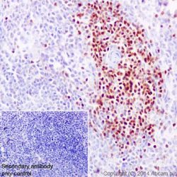
آنتی بادی LEF1 (Lymphoid Enhancer-Binding Factor 1) (EP310)
Name: Lymphoid Enhancer-Binding Factor 1 Antibody clone EP310
Description and applications: LEF1 is a nuclear protein of 42 kDa of molecular weight that is codified by the gen LEF1 located on the chromosomal region 4q25. It belongs to a family of regulatory proteins that share structural homology with high mobility group proteins (HMG1). LEF1 is expressed in pre-B and pre-T lymphocytes, where, through the Wnt signaling pathway, acts as an essential transcription factor for the proliferation and survival of these cells. The activations of the Wnt pathway leads to the accumulation of β-catenin and, eventually, to the gene transcription involved in the survival and cell cycle. The sustained activation with overexpression of LEF1, as it occurs in the chronic ymphoid leukemia, has been proven to play an important role in the molecular carcinogenesis, where its inhibition in experimental animal models leads to apoptosis of the neoplastic cells. In normal lymphoid tissues, the nuclear staining of LEF1 is observed to be predominant in T cells of the paracortical regions without being detected in the B cells, where their differentiation towards mature elements and plasmocytes is accompanied by the loss of the expression of LEF1.
In lymphoid neoplasias, the presence of over 10% of stained tumor cells is considered as positive staining and LEF1 is a marker with high specificity for the diagnosis of chronic lymphoid leukemia (CLL)/small lymphocytic lymphoma, including both the CD5 positive and CD5 negative cases if the cell morphology is concordant. The staining of LEF1 is in direct correlation with the expression of ZAP70 and implies a more favorable prognosis for the neoplasia without observing a correlation with the expression of CD38, the deletion of p53 or the trisomy 12. Nonetheless, and in probable relation with the sensitivity and specificity of the methods and antibodies used for the detection of LEF1, in other lymphomas, variable positive results have been obtained, so that the antibody has focal staining in up to 50% of the high grade follicular lymphomas and 40% of the diffuse large B-cell lymphomas, of which 18% are associated with the transformation of CLL in the Richter’s syndrome. In this later case, the staining is usually more intense in the areas with more atypia. Although most of publications confirm the negativity of the marginal and mantel cell lymphomas, more recent studies have proven focal staining in isolated cases. Additionally, and as LEF1 offers simultaneous staining in the reactive T lymphocytes often trapped within the clonal proliferative process, the correlation of the staining of LEF1 with other T marker as CD3, as well as other specific markers, is very advisable. LEF1 expression in other neoplasias, such as colon or pancreatic adenocarcinomas, has been mentioned in
isolated studies.Composition: Anti-human LEF1 rabbit monoclonal antibody purified from serum and prepared in 10mM PBS, pH 7.4, with 0.2% BSA and 0.09% sodium azide
Intended use: Immunohistochemistry (IHC) on paraffin embedded tissues. Not tested on frozen tissues or Western-Blotting
-
 کیت های لنفوم
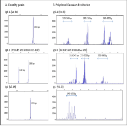
IgH Rearrangements Molecular Analysis Kit
Kit characteristics
Contains an amplification mix for each one of the segments FR1-JH, FR2-JH and FR3-JH, and an internal
control for verifying DNA quality.The amplification mixtures are in “monotest” form in colour-identified PCR tubes of 0.2 – 0.5 ml.
All the mixtures include primers labelled at the 5’ end with 6-FAM fluorochrome, which allows automatic
analysis of fragments by capillary electrophoresis using GeneScan.All amplifications can be carried out in the thermocycler using a single programme.
The enzyme Phire® Hot Start II DNA Polymerase is supplied in the kit.
Positive controls for clonal and polyclonal DNA are included.
-
 کیت های لنفوم
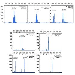
IgK-IgL Rearrangements Molecular Analysis Kit
Kit characteristics
Contains an amplification mix for each one of the segments IgK (Vκ-Jκ, Vκ-Kde and intronRSS-Kde
rearrangements), IgL (Vλ-Jλ), and an internal control for verifying DNA quality (CI).
The amplification mixtures are in “monotest” form in colour-identified PCR tubes of 0.2 ml.
All the mixtures include primers labelled at the 5’ end with 6-FAM fluorochrome, which allows automatic
analysis of fragments by capillary electrophoresis (ABI PRISM® 310 Genetic Analyzer, AB 3130, 3130xl,
3500 y 3500xL Genetic Analyzers).
All amplifications can be carried out in the thermocycler using a single programme.
The enzyme Phire® Hot Start II DNA Polymerase is supplied in the kit.
Positive controls for clonal and polyclonal DNA are included.REAGENT COMPONENTS QUANTITY
Amplification mixtures IgK-A (6-FAM) 20 PCR tubes blue (46 μl)
(monotest in tubes of 0.2) (mix of oligonucleotides from regions Vκ
and Jκ of IgK gene, dNTP and buffer solution)IgK-B (6-FAM) 20 PCR tubes:orange (46 μl)
(mix of oligonucleotides from regions Vκ,
intron-RSS, and Kde of IgK gene, dNTP and buffer solution)IgL (6-FAM) 20 PCR tubes green (46 μl)
(mix of oligonucleotides from regions Vλ-
Jλ of IgL gene, dNTP and buffer solution)Internal control mix (CI) (6-FAM) 20 PCR tubes yellow (46 μl)
(oligonucleotides specific for exon 5 of p53
gene, dNTP and buffer solution)DNA polymerase Phire® Hot Start II 2 x 60 μl
DNA Polymerase*Clonal IgK-IgL positive control DNA 1 x 50 μl
(50 μg/ml)Positive control DNAs Polyclonal positive control DNA 1 x 50 μl
(100 μg/ml) -
 کیت های لنفوم
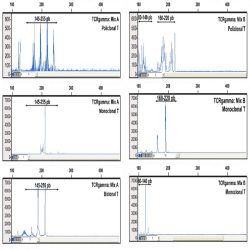
TCR Beta Rearrangements Molecular Analysis Kit
Kit characteristics
Contains two reaction mixtures (Mixes A and B) for amplification of the Vß-Jß region and a mixture (Mix C) for
Dß-Jß region of the TCRß gene with an internal amplification control for checking the quality of the DNA.The amplification mixtures are in “monotest” form in colour-identified PCR tubes of 0.2 – 0.5 ml.
All the PCR mixtures include primers marked at their 5’ end with 6-FAM fluorochrome, allowing automatic
analysis of fragments by capillary electrophoresis using GeneScan.All amplifications can be carried out in the thermocycler using a single programme.
The enzyme Phire® Hot Start II DNA Polymerase is supplied in the kit.
Positive controls for clonal and polyclonal DNA are included.
-
 کیت های لنفوم
TCR Gamma Rearrangements Molecular Analysis Kit
Kit characteristics
Contains two multiplex reaction mixtures (Mixes A and B) for the V-J region of the TCRgamma gene with an
internal amplification control for checking the quality of the DNA.The amplification mixtures are in “monotest” form in colour-identified PCR tubes of 0.2 – 0.5 ml.
All the PCR mixtures include primers marked at their 5’ end with 6-FAM fluorochrome, allowing automatic
analysis of fragments by capillary electrophoresis using GeneScan.All amplifications can be carried out in the thermocycler using a single programme.
The enzyme Phire® Hot Start II DNA Polymerase is supplied in the kit.
Positive controls for clonal and polyclonal DNA are included.
-
 ایمونو هیستوشیمی
EDTA Buffer 10X pH8
Product Composition: 10mM EDTA pH8.0.
Intended Use: Product for in vitro diagnostic use
Recommendations for Use:
Before use: The EDTA solution is 10x concentrated and therefore before use it should be diluted in a 1:10 range with distilled or deionized water (one part EDTA buffer and 9 parts water).
-Cut and mount sections on charged slides or silane coated.-Dewaxing (using xylene or substitute) and rehydrate the tissue sections thru decreasing series of alcohol. Then follow with the step 3 and 4.
-Unmasking: various alternative methods could be used to carry out the antigen retrieval
process using the diluted EDTA buffer-Remove the sections and place them into the usual washing buffer (recommended TBS / TBS-Tween pH 7.5 – reference MAD-004077R)
-Proceed with the immunostaining protocol as usual.
-
-
 ایمونو هیستوشیمی
Tris-EDTA Buffer 10X pH9
Product Composition: TRIS BASE and EDTA pH 9.00
Intended Use: Product for in vitro diagnostic use
Recommendations for Use:
Before use: The TRIS EDTA solution is 10x concentrated and therefore it must be diluted in a
1:10 range with distilled or deionized water (one part TRIS EDTA buffer and 9 parts water).
1. – Cut and mount sections on either charged or silane coated slides.
2A. – In case of using an automated PT Module for simultaneous dewaxing, rehydrating and antigen (HIER) retrieval, follow the instructions enclosed in the equipment data sheet. Basically, fill the tanks with 1.5 litres of diluted, ready-to-use buffer solution, place the slides without dewaxing into the racks and then immerse them into the buffer solution. Program the PT Module for a cycle of antigen retrieval of 20min at 95ºC and then cool to room temperature. Then follow with the step 4.
2B. – In case of not using the PT Module for HIER continue with dewaxing (using xylene or substitute) and rehydration of the tissue sections through decreasing series of ethanol. Then follow with the step 3 and 4.3. – Unmasking: different alternative methods could be used to carry out the antigen retrieval process using the diluted TRIS EDTA buffer:
3.1 Unmasking in microwave: Place the sections in the diluted solution of TRIS EDTA (Coplin jar or other container with a volume as large as the laboratory considers) and bring the microwave oven to 700 watts (watt) for 10-15 minutes). Cool to room temperature.
3.2 Unmasking in pressure cooker: Fill the pressure cooker with 1-2 litres of diluted TRIS EDTA buffer. While hydrating the slides, heat the solution avoiding boiling. Then immerse the sections in the preheated buffer and close the pressure cooker. Wait until the pressure rises to the second notch and run from here 1-2 minutes (depending on the antigen unmasking). Remove and cool the pot (you can speed up the process placing the pot under running water). Open when there is no pressure and cool the slides inside of the pot for 20-30 minutes.
4. – Remove the sections and place them into the usual washing buffer (recommended TBS / TBS-Tween pH 7.5 – reference MAD-004077R)
5. – Proceed with the immunostaining protocol as usual -
 ایمونو هیستوشیمی
Citrate Buffer 10X PH6
Product Composition: Sodium- CITRATE pH6.0.
Intended Use: Product for in vitro diagnostic use
Recommendations for Use:
Before use: The CITRATE solution is 10x concentrated and therefore it must be diluted in a 1:10 range with distilled or deionized water (one part CITRATE buffer and 9 parts water).
1. – Cut and mount sections on either charged or silane coated slides.
2A. – In case of using an automated PT Module for simultaneous dewaxing, rehydrating and antigen (HIER) retrieval, follow the instructions enclosed in the equipment data sheet. Basically, fill the tanks with 1.5 litres of diluted, ready-to-use buffer solution, place the slides without dewaxing into the racks and then immerse them into the buffer solution. Program the PT Module for a cycle of antigen retrieval of 20min at 95ºC and then cool to room temperature. Then follow with the step 4.
2B. – In case of not using the PT Module for HIER continue with dewaxing (using xylene or substitute) and rehydration of the tissue sections through decreasing series of ethanol. Then follow with the step 3 and 4.3. – Unmasking: different alternative methods could be used to carry out the antigen retrieval
process using the diluted CITRATE buffer:
3.1 Unmasking in microwave: Place the sections in the diluted solution of CITRATE (Coplin jar or other container with a volume as large as the laboratory considers) and bring the microwave oven to 700 watts (watt) for 10-15 minutes). Cool to room temperature.
3.2 Unmasking in pressure cooker: Fill the pressure cooker with 1-2 litres of diluted CITRATE buffer. While hydrating the slides, heat the solution avoiding boiling. Then immerse the sections in the preheated buffer and close the pressure cooker. Wait until the pressure rises to the second notch and run from here 1-2 minutes (depending on the antigen unmasking). Remove and cool the pot (you can speed up the process placing the pot under running water). Open when there is no pressure and cool the slides inside of the pot for 20-30 minutes.
4. – Remove the sections and place them into the usual washing buffer (recommended TBS / TBS-Tween pH 7.5 – reference MAD-004077R)
5. – Proceed with the immunostaining protocol as usual.
برای تماس کلیک کنید
سما تشخیص آریا ، عرضه کننده متنوع ترین و کامل ترین محصولات کیت های آزمایشگاهی