-
 آنتی بادیهای ایمونوهیستوشیمی
آنتی بادی Epithelial Specific Antigen (BER-EP4)
Name: Epithelial Specific Antigen-Clone Ber-EP4
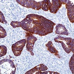
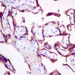
Description and aplications: This antibody reacts with two glycoproteins of 34 and 49 kD molecular weight present on the surface and in the cytoplasm of all epithelial cells except the surface layer of the squamous epithelium, hepatocytes and parietal cells of the stomach. This antibody is specific to HEA125 equivalent. The antibody shows a broad pattern of reactivity with human epithelial tissues like simple epithelium, basal layer of the stratified squamous non-keratinized epithelia and epidermis. In contrast to other known anti-epithelial antibodies, this antibody does not mark the mesothelial cells and only a small number of mesotheliomas stained positively. This antibody does not react with nervous, glial, muscle, mesenchymal or lymphoid tissue. Due to the labile nature of the epitope in tissues fixed in buffered formalin and embedded in paraffin, negative staining results should be considered with caution.
Composition: anti-Ber-EP4 mouse monoclonal antibody obtained from supernatant culture and prediluted in a tris buffered solution pH 7.4 containing 0.375mM sodium azide solution as bacteriostatic and bactericidal.
Immunogen: MCF-7 human breast carcinoma cell line.
-
 آنتی بادیهای ایمونوهیستوشیمی
آنتی بادی Epstein-Barr Virus / LMP1 (CS1-4)
Name: Epstein-Barr Virus / LMP1 (Clone CS1-4)
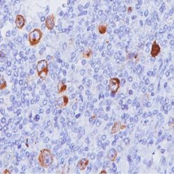
Description and applications:The antibody strongly reacts with EBV-positive lymphoblastoid cell lines and EBV infected B cell immunoblasts in infectious mononucleosis. It also reacts with 25 to 50 per cent of EBV associated undifferentiated nasopharyngeal carcinomas and with Reed Sternberg cells in approximately 90% of EBVassociated Hodgkin’s disease cases. The cocktail recognizes distinct epitopes on the hydrophilic carboxyl region of LMP which is exposed to the cytosol.
Composition:Anti-human Epstein-Barr Virus / LMP1 mouse monoclonal antibody purified from serum and prepared in 10mM PBS, pH 7.4, with 0.2% BSA and 0.09% sodium azide.
Immunogen: EBV-encoded recombinant latent membrane protein.
-
 آنتی بادیهای ایمونوهیستوشیمی
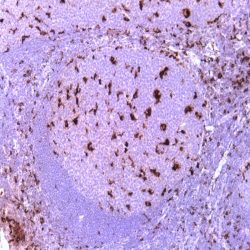
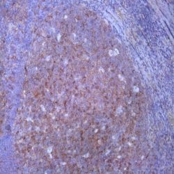
آنتی بادی Fascin (FCN01)
Name: Fascin (Clone FCN01)
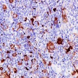
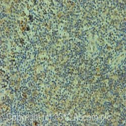
Description and applications:Anti-Fascin is a very sensitive marker for ReedSternberg cells and variants in nodular sclerosis, mixed cellularity, and lymphocyte depletion Hodgkin’s disease. It is uniformly negative in lymphoid cells, plasma cells and myeloid cells. Anti-Fascin is positive in dendritic cells. This marker may be helpful in distinguishing between Hodgkin’s disease and nonHodgkin’s lymphoma in difficult cases. Also, the lack of expression of Fascin in the neoplastic follicles in follicular lymphoma can be helpful in distinguishing these lymphomas from reactive follicular hyperplasia in which the number of follicular dendritic cells is normal or increased. Anti-Fascin has been suggested as a prognostic marker in neuroendocrine neoplasms of the lung, as well as ovarian cancer.
Composition: Anti-human Fascin mouse monoclonal antibody purified from serum and prepared in 10mM PBS, pH 7.4, with 0.2% BSA and 0.09% sodium azide.
-
 آنتی بادیهای ایمونوهیستوشیمی
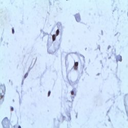
آنتی بادی Gastrin (Polyclonal)
Name: Rabbit anti-human Gastrin Antibody
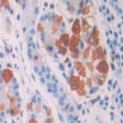
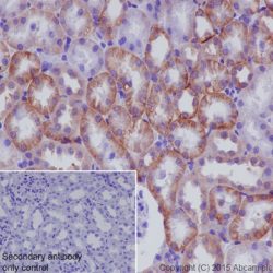
Description and applications: Gastrin is a 17 amino acid peptide hormone and neurotransmitter widely distributed throught the gastrointestinal (GI) tract and the central nervous system (CNS). Antibodies that react specifically with gastrin may be used to study the differential tissue expression and intracellular and subcellular localization of gastrin in neuroendocrine cells of the gastrointestinal tract, and in the CNS. Antibodies to gastrin are also useful for the identification and detection of gastrin in normal and neoplastic tissue.
Available Volume: 3ml / 7ml / 12ml / Concentrated
Composition: anti-human Gastrin rabbit polyclonal antibody purified from serum and prepared in 10mM PBS, pH 7.4, with 0.2% BSA and 0.09% sodium azide
Intended use: Immunohistochemistry (IHC) on paraffin embedded tissues. Not tested on frozen tissues or Western-Blotting
Immunogen: Synthetic peptide corresponding to Nterminus of Gastrin 1.
-
 آنتی بادیهای ایمونوهیستوشیمی
آنتی بادی GH (Growth Hormone) (EP267)
Name: Rabbit anti-human GH (Growth Hormone) Monoclonal Antibody clone EP267
Description and applications: Growth hormone (GH or hGH), also known as somatotropin or somatropin, is a peptide hormone that is produced and secreted by somatotrophs of the anterior pituitary gland. GH exerts a wide variety of biological actions in many different tissues and cell types. The actions of GH at the cellular level can be divided into three categories: those affecting mitogenesis, differentiation, and metabolism.The GH antibody specifically labels somatotrophs in pituitary in normal tissues. It is useful in classification of pituitary tumor.
Composition: Anti-human GH (Growth Hormone) rabbit monoclonal antibody purified from serum and prepared in 10mM PBS, pH 7.4, with 0.2% BSA and 0.09% sodium azide
Immunogen: A synthetic peptide corresponding to residues of human Growth Hormone protein
-
 آنتی بادیهای ایمونوهیستوشیمی
آنتی بادی GLUT 1 (Polyclonal)
Name: glut-1 Antibody
Description and applications: Glucose transporter type I (GLUT1), a prototype member of GLUT super family, reacts with a 55 kD protein, is a membrane-associated erythrocyte glucose transport protein. It is a major glucose transporter in the mammalian blood-brain barrier, and also mediates glucose transport in endothelial cells of the vasculature, adipose tissue and cardiac muscle. GLUT1 is detectable in many human tissues including those of colon, lung, stomach, esophagus, and breast. GLUT1 is overexpressed in malignant cells and in a variety of tumors that include the breast, pancreas, cervix, endometrium, lung, mesothelium, colon, bladder, thyroid, bone, soft tissues, and oral cavity. Immuohistochemical detection of GLUT1 can discriminate between reactive mesothelium and malignant mesothelioma. Anti-GLUT1 with anti-Claudin1, and anti-EMA are “perineurial” markers in diagnosis of perineuriomas. Anti-GLUT1 is also useful in distinguishing benign endometrial
hyperplasia from atypical endometrial hyperplasia and adenocarcinoma. GLUT1 expression has been associated with increased malignant potential, invasiveness, and a poor prognosis in general. Expression of GLUT1 is a late event in colorectal cancer and expression in a high proportion of cancer cells is associated with a high incidence of lymph node metastasesComposition: anti-human GLUT 1 rabbit polyclonal antibody purified from serum and prepared in 10mM PBS, pH 7.4, with 0.2% BSA and 0.09% sodium azide
-
 آنتی بادیهای ایمونوهیستوشیمی
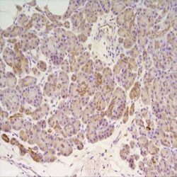
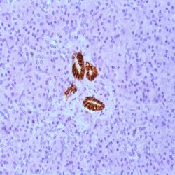
آنتی بادی Glutamine Synthetase (GS-6)
Name: Glutamine Synthetase (Clone GS-6)
Description and applications:Glutamine synthetase (GS) catalyzes the synthesis of glutamine from glutamate and ammonia in the mammalian liver. In normal liver, GS expression is seen in pericentral hepatocytes, but not in mid-zonal or periportal hepatocytes. Glutamine, the end product of GS activity, is the major energy source of tumor cells. Based on findings from experimental hepatocarcinogenesis, GS positive tumor cells are believed to be derived from GS positive hepatocytes. Thus, anti-GS has been suggested as a marker for hepatocellular carcinoma (HCC). GS immunoreactivity has been seen in a majority of HCC (37 of 53 cases, 70%), including 7 of 10 cases of early HCC (70%) and 12 of 22 (59%) for low grade HCC. In nonmalignant nodules, GS overexpression was only seen in 3 high grade dysplastic nodules (HGDN,13.6%). In these cases, GS overexpression was restricted to 11.5%-50% of hepatocytes, whereas in HCC the majority of cases (28 of 53, 53%), including early HCC (60%), showed diffuse immunostaining (>50% tumor cells). Overall, the sensitivity, specificity, and positive and negative predictive values of anti-GS for HCC detection were 69.8%, 94.2%, 92.5%, and 75.4%, respectively. A panel composed of antibodies against HSP70, GPC3, and GS has been proposed to be very useful in distinguishing between dysplastic and early malignant hepatocellular nodules arising in cirrhosis. The “all positive” phenotype is restricted to approximately half of early HCC to well-differentiated HCC but has never been reported in dysplastic lesions, whereas the reverse phenotype, “all negative”, has been shown to be a feature of the majority of HGDN and of all low grade dysplastic nodules.
Composition: Anti-human Glutamine Synthetase mouse monoclonal antibody purified from serum and prepared in 10mM PBS, pH 7.4, with 0.2% BSA and 0.09% sodium azide.
-
 آنتی بادیهای ایمونوهیستوشیمی
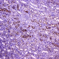
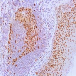
آنتی بادی PAX-2 (EP235)
name: PAX-2 Antibody clone EP235
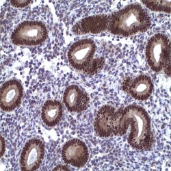
Description and aplications: PAX2 is a member of the paired box family of transcription factors, which is required for development and proliferation of the kidney, brain, and müllerian organs. PAX2 genes contain a highly conserved DNA sequence within the paired box region, which encodes a DNA-binding domain, enabling PAX proteins to bind the promoters of specific genes to transcriptionally regulate their expression. Defects in PAX2 gene are related with the renal coloboma syndrome (RCS) (also known as papillorenal syndrome) which is a condition that primarily affects kidney (renal) and eye development. PAX2 is specifically expressed in the developing central nervous system, eye, ear, and urogenital tract, and is essential for the development of these organs. In normal adult tissues PAX2 was mainly detected in the urogenital system, including kidney, uretericepithelium, fallopian tube epithelium, ovary and uterus. In tumors, PAX2 has been detected in renal cell carcinomas, Wilms’ tumors, nephrogenic adenomas and papillary serous carcinoma of the ovary. For these reasons, PAX2 has been used as a marker for the identification of renal cell carcinoma and ovarian carcinoma by immunohistochemistry. It has been also suggested as a useful tool in diagnosis and classification of hyperplastic endometrial epithelial proliferations
Composition: anti-human PAX2 rabbit monoclonal antibody purified from ascites fluid by chromatography. Prepared in 10mM PBS, pH 7.4, with 0.2% BSA and 0.09% sodium azide
-
 آنتی بادیهای ایمونوهیستوشیمی
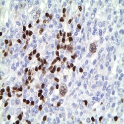
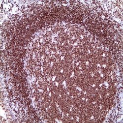
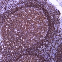
آنتی بادی Pax-5 (MX017)
Name: PAX-5 Antibody clone MX017
Description and applications: The PAX-5 gene is essential for B-cell differentiation. There are at least four isoforms, of which PAX-5a has been most studied. PAX-5 encodes the 50 kDa B-cell specific activator protein, BSAP. PAX-5 is expressed by pro-, pre-, and mature B-cells, but is downregulated during terminal differentiation of plasma cells. PAX-5 influences the expression of other B-cell specific genes, including CD19 and CD20 and CD79a, preceding the expression of CD20. PAX-5 is silenced at the plasma cell stage under the influence of Blymphocyte-induced maturation protein-1 (PRDM1). Pax-5 is consistently expressed in mature B cell lymphomas. It is useful to distinguish between classical Hodgkin lymphoma (+) and ALCL (-). In Hodgkin lymphoma, the neoplastic cells show a weaker staining compared with the accompanying B cells. Occasionally it expressed in acute myeloid leukemias especially those with t (8; 21). CD20 negative B-cell lymphomas of patients treated with anti-CD20 monoclonal antibodies, maintain PAX5 staining the antibody representing a good tool to confirm their cell of origin. Pax-5 expression was detected in hyperplastic mesonephric remains, epididymitis, neurons of the adult nervous tissue, small cell carcinomas, Merkel cell tumor and other neuroendocrine tumors.
Composition: anti-human PAX5 mouse monoclonal antibody purified from serum and prepared in 10mM PBS, pH 7.4, with 0.2% BSA and 0.09% sodium azide
-
 آنتی بادیهای ایمونوهیستوشیمی
آنتی بادی Alpha-1-Antichymotrypsin (Polyclonal)
Name: Alpha-1-Antichymotrypsin (Polyclonal)
Description and applications: This antibody reacts with the human alpha-1- antichymotrypsin, a surfactant protein in acute phase of the inflamation, plus a serpin that inactivates preferably the chymotrypsin, the cathepsin G and the chymase. As an inhibitor protein of serine protease, it is closely related to the neuritic plaques in the Alzheimer’s disease and in human and monkey brains with normal aging signs. The regulation of serine proteases and their inhibitors have an important function in the neuromuscular pathology differential diagnosis. The alpha-1-antichymotrypsin binds primarily to the prostate specific antigen (PSA), a serine protease similar to the chymotrypsin, to form a complex. Furthermore, this antibody can be used to identify hematopoietic cells of the monocyte-macrophage series and neoplasias derived from them.
Composition: anti-human Alpha-1-Antichymotrypsin rabbit polyclonal antibody purified from serum and prepared in 10mM PBS, pH 7.4, with 0.2% BSA and 0.09% sodium azide
Clone: Polyclonal
Ig isotype: Rabbit IgG
IHC positive control: Macrophages in the tonsils.
Visualization: Cytoplasmic.
-
 آنتی بادیهای ایمونوهیستوشیمی
آنتی بادی C3d Complement Component (Polyclonal)
Name: C3d Antibody
Description and applications: This antibody recognises the C3d fragment of the complement. Component C3 can be activated through both classical and alternative complement pathways. Among the different complement proteins, C3 and C4 opsonins have a protective thioester radical. This molecule enables C3 (and C4) complement fragments to form covalent bonds when activated by the target molecule, generating C3b. The proteolytic cleavage of C3b generates the C3d fragment, which remains covalently attached to the target structure, while the C3c fragment is released. The C3d fragment is stable and it can be identified in paraffin-embedded biopsies. Under normal conditions, renal glomerular capillaries are stained. In transplants, the identification of diffuse deposits of C3d and C4d in renal peritubular capillaries or cardiac capillaries supports the diagnosis of acute humoral rejection of the graft. Deposits of C3d have been identified in grafts with chronic rejection but its significance has yet to be clarified. Deposition of C3d on paraffin-embedded biopsies from skin inflammatory pathologies has been demonstrated by different authors. Magro and Dyrsen identified prominent granular C3d deposits along the dermoepidermal junction for all discoid lupus erythematosus (20/20) and systemic LE (5/5) cases. In systemic LE, vascular C3d or C4d deposits were shown. All cases of urticarial (5), leukocytoclastic (6), and lymphocytic (1) vasculitis exhibited prominent mural C3d and C4d deposits in vessels, whereas Henoch-Schönlein purpura (10/10) showed only C3d deposits. Pfaltz et al found deposits of C3d at the epidermal basement membrane in 97% (31/32) of bullous pemphigoid cases.
Composition: anti-human C3d Complement Component rabbit polyclonal antibody purified from serum and prepared in 10mM PBS, pH 7.4, with 0.2% BSA and 0.09% sodium azide
IHC positive control: Kidney tissue section with acute humoral rejection.
-
 آنتی بادیهای ایمونوهیستوشیمی
آنتی بادی CA 19-9 (121SLE)
Name : Antibody Ca19-9 clone 121SLE
Description and applications: CA19-9 antigen is highly expressed in gastrointestinal (gastric, pancreatic, and colonic) adenocarcinomas and salivary gland mucoepidermoid carcinomas. Anti- CA19-9 antibody is usually not reactive with breast, kidney, and prostate carcinomas. Anti-human CA19-9 mouse monoclonal antibody purified from serum and prepared in 10mM PBS, pH 7.4, with 0.2% BSA and 0.09% sodium azide
Composition:Anti-human CA19-9 mouse monoclonal antibody purified from serum and prepared in 10mM PBS, pH 7.4, with 0.2% BSA and 0.09% sodium azide.
-
 آنتی بادیهای ایمونوهیستوشیمی
آنتی بادی PAX-8 (MD-50 also known as MRQ-50)
Name: Pax-8 Antibody clone MD-50 also known as MRQ-50
Description and applications:PAX8 antibody recognizes a protein of 62kDa, identified as PAX8. It is a member of the paired box (PAX) family of transcription factors. This nuclear protein is involved in thyroid follicular cell development and expression of thyroid-specific genes. Mutations in this gene have been associated with thyroid dysgenesis, thyroid follicular carcinomas, and atypical thyroid adenomas. PAX8 is expressed in the normal thyroid follicular cells and associated carcinomas. As a marker of mesonephric and müllerian organs, ovarian and renal normal and neoplasms are frequently positive. For that, non-ciliated mucosal cells of the fallopian tubes, and simple ovarian inclusion cysts and normal ovarian surface epithelial cells are positive. PAX8 is expressed in a high percentage of ovarian serous, endometrioid, and clear cell carcinomas, but only rarely in primary ovarian mucinous adenocarcinomas. PAX8 antibody may be used as an additional immunohistochemical marker for renal epithelial tumors as it is expressed by all the segments of the nephron and the great majority of renal tumours including sarcomatoid carcinomas. Thymic and parathyroid carcinomas and isolated cases of urothelial carcinomas, sex-cord stromal tumours of the ovary or squamous carcinomas of the lung have showed weak and focal positivity.
Composition:Anti-human PAX-8 mouse monoclonal antibody purified from serum and prepared in 10mM PBS, pH 7.4, with 0.2% BSA and 0.09% sodium azide
-
 آنتی بادیهای ایمونوهیستوشیمی
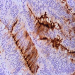
آنتی بادی Cadherin 17 (SP183)
Name:Cadherin 17 Antibody clone SP183
Description and applications: Cadherins represents a big family of Ca2+ dependent transmembrane cell adhesion glycoproteins that interact homophilically with each other and thus participate in the heterologous cell selection. They play a key role in the embryonic development and in the maintenance of the normal structure in adult tissues. Molecular defects in the structure of cadherins represent important pathogenic aspects of many diseases, including cancer. The cadherin 17 gene (CDH17) is located on the chromosome region 8q22.1 that encodes a protein of 832 amino acids and approximately 92kDa of molecular weight. Cadherin 17, also known as human peptide transporter 1 (HPT-1) or liver-intestine cadherin (LICadherin), is located in the lateral contact areas of the intestinal epithelial cells, being undetectable in the stomach, kidney, lung or liver, among other organs. It
features many structural similarities with the kidney specific cadherin, both belonging to the 7D-cadherin family. In tumors, the great specificity of cadherin 17 to identify the primary or metastatic colorectal adenocarcinomas has been demonstrated. Less frequently, cadNherin 17 has been described in gastroesophageal junction adenocarcinomas or adenocarcinomas with gastric origin. In the few available studies, cadherin 17 has shown staining in all well-differentiated neuroendocrine tumors (carcinoid tumors) originated in the small intestine and in the appendix. Up to 80% of the primary pancreatic adenocarcinomas and only half of their metastases are usually positive. Up to 50% of cholangiocarcinomas show membrane staining against cadherin 17, showing greater sensitivity than other markers such as villin, CDX2 or SATB2, which can be used to refine the diagnosis. Cadherin 17 shows along with SATB2 greater sensitivity in the detection of medullar carcinomas of the large intestine that are usually negative for CDX2 and cytokeratins 7 and 20.Composition: Anti-human Cadherin 17 rabbit monoclonal antibody purified from serum and prepared in 10mM PBS, pH 7.4, with 0.2% BSA and 0.09% sodium azide
-
 آنتی بادیهای ایمونوهیستوشیمی
آنتی بادی CD3 (EP41)
Name: CD3 Antibody clone EP41
Description and applications: CD3 (Cluster of Differentiation 3) is a complex of proteins that associates directly with the T cell antigen receptor (TCR). CD3 is composed of five invariant polypeptide chains that associate to form three dimers. The five invariant chains of CD3 are labeled gamma, delta, epsilon, zeta, and eta. The CD3 is involved in T cell development and survival. It is expressed on T cells in thymus, peripheral lymphoid tissue, blood and bone marrow. CD3 is a commonly used marker for identification of T cell and T cell derived malignancies. This CD3 antibody has been validated by the 9th International Conference on Human Leukocyte Differentiation Antigens (HLDA9).
Composition: Anti-human CD3 rabbit monoclonal antibody purified from serum and prepared in 10mM PBS, pH 7.4, with 0.2% BSA and 0.09% sodium azide
Intended use: Immunohistochemistry (IHC) on paraffin embedded tissues. Not tested on frozen tissues or Western-Blotting
Species reactivity: In vitro diagnostics in humans. Not tested in other species
-
 آنتی بادیهای ایمونوهیستوشیمی
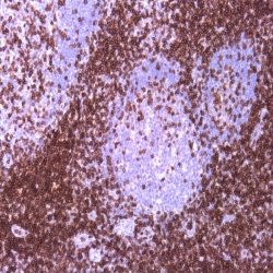
آنتی بادی CD45RA (SPM504)
Name: CD45RA Monoclonal Antibody clone SPM504
Description and applications: This antibody reacts with the CD45RA isoform with a molecular mass of 220 kD. CD45RA is present in peripheral blood B cells, follicular mantle and centre, medullary thymocytes, monocytes, and mature T cells that have not been modulated by antigenic contact. In this regard, and after immune processing in the thymus, T cells are released into peripheral blood to colonize secondary lymphoid organs, including spleen, lymph nodes, and mucosa-associated lymphoid tissue (MALT). These lymphocytes, not yet activated in vivo by antigens (naive cells), are phenotypically characterized by the co-expression of the highest molecular mass CD45 isoforms, CD45RAC and CD45RA, and CD62L (peripheral lymph node homing receptor), a decisive molecule for the fixation of cells in thymus-dependent areas of secondary lymphoid organs. When these cells are stimulated by its specific antigen expressed by the corresponding antigen-presenting cells and are transformed into effector T cells, either CD4- or CD8-positive or memory T cells, CD45RA and CD62L expression disappears, the former being replaced by the lower molecular mass CD45R0 isoform, and the latter, by various type 1 and type 2 integrins and APO-1/Fas receptor (CD95), an apoptosis inducer. The CD45 molecule, also called leukocyte common antigen, is a membrane glycoprotein that occupies up to 10% of the surface of cells expressing it. In mammals, its homology varies markedly between the intracytoplasmic region (greater than 90%) and the extracellular region (only 35%, although with similar domain organization). Structurally, there are multiple CD45 isoforms determined by the different possible ways of joining exons 4, 5, and 6 during the synthesis of the mRNA encoding the extracellular domain of the protein. These isoforms are referred to as A, B, and C, using the RABC terminology for the bigger isoform, which includes all three exons, RA and RB when the exonic expression is restricted to one of these exons, and RO to designate the smaller molecule, which lacks all three exons. The sequence of these three exons includes multiple oxygen-linked glycosylation sites that can be variably modified by sialic acid. This, on the one hand, causes the molecular mass of the different isoforms to vary between 180 kD (RO) and 240 kD (RABC); on the other hand, it gives these molecules remarkable differences in terms of their shape and anionic charge. The rest of CD45’s extracellular domain shows marked nitrogen-linked glycosylation and contains a cysteine-rich region followed by three amino acid repeats analogous to fibronectin type III domain. CD45’s high degree of glycosylation, attributable to both sialic acid and oligosaccharides on the variable domains and N-glycoconjugates on the constant extracellular domain, plays an important role in the functional activity of the molecule, as demonstrated by the interaction between CD45RO of T cells and B cells expressing CD22 or the mannose hybrids on CD45 in the development of immature thymocytes. The rest of the CD45 molecule consists of a single transmembrane domain followed by a long intracytoplasmic tail containing two tandem-placed identical domains (D1 and D2) with protein-tyrosine phosphatase (PTPase) homology. Of these domains, only D1 possesses enzymatic activity capable of rescuing TCR signal activation in CD45-deficient cell lines. The function of D2 is hardly understood, although it seems to contribute to the molecule’s intracytoplasmic stability. Although in in vitro models the extracellular domain of CD45 is not essential for its intracytoplasmic function, the exonic transcription of this domain is strongly regulated depending on the cell type that expresses it, its development, and its degree of activation, thus playing an important functional role in vivo.The CD45RA epitope recognized by the MB1 antibody is useful for the study of B-cell lymphomas; however, as some of them are not positive against it, it is advisable to use it as part of a large panel of antibodies. Because the epitope recognized by MB1 may be altered by prolonged formol fixation, and in order to ensure a correct interpretation of results, it is appropriate to check that at least some normal B cells in the histological section are stained. The antibody shows no reactivity against cortical thymocytes, immature T cells, and activated B cells. This antibody reacts against human tissue. In other species, it has not been tested.
Composition: Anti-human CD45RA mouse monoclonal antibody purified from serum and prepared in 10mM PBS, pH 7.4, with 0.2% BSA and 0.09% sodium azide
-
 آنتی بادیهای ایمونوهیستوشیمی
آنتی بادی CD68 (KP-1)
Name: CD68 Monoclonal Antibody clone KP-1
Description and aplications: Anti-CD68 marks cells of monocyte/macrophage lineage. This antibody is capable of staining monocytes, Kupffer cells, osteoclasts, NK cells, granulocytes and their precursors; lymphomas are negative or show a few granules. This antibody may be useful for the identification of myelomonocytic and histiocytic tumors. Anti-CD68 may help to distinguish malignant fibrous histiocytoma from other pleomorphic sarcomas. However, since this detects a formalinresistant epitope that may be associated with lysosomal granules, other lysosome-rich cells may also stain.
Composition: Anti-human CD68 mouse monoclonal antibody purified from serum and prepared in 10mM PBS, pH 7.4, with 0.2% BSA and 0.09% sodium azide
-
 آنتی بادیهای ایمونوهیستوشیمی
آنتی بادی Cdk4 (DCS-35)
Name: Anti-human CDK4 Mouse Monoclonal Antibody clone DCS-35
Description and aplications: Cell cycle progression is controlled in part by a family of cyclin proteins and cyclin dependent kinases (Cdks). Cdk proteins work in concert with the cyclins to phosphorylate key substrates involved in each phase of cell cycle progression.
Another family of proteins, Cdk inhibitors, also plays a role in regulating the cell cycle by binding to cyclin-Cdk complexes and modulating their activity. Several Cdk proteins have been identified, including Cdk2-Cdk8, PCTAIRE-1-PCTAIRE-3, PITALRE and PITSLRE. Cdk4, in complex with D-type cyclins, is thought to regulate cell growth during the G1 phase of the cell cycle. This association with a D-type cyclin upregulates Cdk4 activity, whereas binding to the Cdk inhibitor p16 downregulates Cdk4 activity. Activation of the Cdk4-cyclin complexes requires phosphorylation on a single threonyl residue of Cdk4, catalyzed by a Cdk-activating protein (CAK).Composition: Anti-human CDK4 mouse monoclonal antibody purified from serum and prepared in 10mM PBS, pH 7.4, with 0.2% BSA and 0.09% sodium azide
Intended use: Immunohistochemistry (IHC) on paraffin embedded tissues. Not tested on frozen tissues or Western-Blotting
-
 آنتی بادیهای ایمونوهیستوشیمی
آنتی بادی CD95 (EP208)
Name: CD95 Monoclonal Antibody clone EP208
Description and aplications: The CD95 (Fas) protein is a cell surface receptor belonging to the tumor necrosis factor (TNF) family that transduces death signaling on engagement by multimeric Fas ligand (CD95L), of which there are eight in its membrane–bound form or in its soluble form resulting from cleavage by a putative metalloproteinase. CD95 is a widely expressed protein. CD95-mediated apoptosis is an essential mechanism for the maintenance of normal tissue homeostasis, and disruption of this death pathway has been associated with a wide range of human diseases, including autoimmune diseases, lymphoproliferative disorders and malignancies. The Fas death system also plays important roles in various apoptosis conditions such as those evoked by irradiation, chemotherapeutic
agents and viral infections. The expression of CD95 serves as a prognostic marker in predicting the outcome of disease progression and treatment in many types of tumors.Composition: Anti-human CD95 rabbit monoclonal antibody purified from serum and prepared in 10mM PBS, pH 7.4, with 0.2% BSA and 0.09% sodium azide
-
 آنتی بادیهای ایمونوهیستوشیمی
آنتی بادی CNPasa (11-5B)
Name: CNPase Monoclonal Antibody clone 11-5B
Description and aplications: This monoclonal antibody recognises two proteins with a molecular mass of 46 kD (CNP1) and 48 kD (CNP2), identified as CNPase isoforms. CNPase (2’,3’-cyclic nucleotide 3′-phosphodiesterase) is expressed at high levels in oligodendrocytes of the central nervous system and in Schwann cells of the peripheral nervous system and it is virtually absent in other cell types. Since CNPase is expressed early in postnatal development, this antibody is useful for the identification of oligodendrocytes and Schwann cells. CNPase activity is decreased in demyelinating diseases such as multiple sclerosis. The CNPase gene is located on chromosome 17q21, very close to the familial breast cancer gene BRCA1. This antibody shows cross-reactivity with monkey, cow, sheep, goat, pig, cat, dog, rabbit, rat, and mouse. It does not cross-react with guinea pig and chicken.
Composition: Anti-human CNPase mouse monoclonal antibody purified from serum and prepared in 10mM PBS, pH 7.4, with 0.2% BSA and 0.09% sodium azide
Intended use: Immunohistochemistry (IHC) on paraffin embedded tissues. Not tested on frozen tissues or Western-Blotting
برای تماس کلیک کنید
سما تشخیص آریا ، عرضه کننده متنوع ترین و کامل ترین محصولات کیت های آزمایشگاهی