-
 آنتی بادیهای ایمونوهیستوشیمی
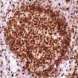
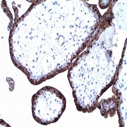
آنتی بادی PU-1 (EP18)
Name: Antibody PU-1
Description and applications: This antibody recognises a protein with a molecular mass of 42 kDa corresponding to PU.1 transcription factor, a member of the Ets family of transcription factors. It is required for the normal development and maturation of multiple hematopoietic lineages. PU.1 is a transcription factor restricted to the hematopoietic system, expressed in the myeloid series and in B cells but absent in mature T cells, the erythroid series, and in non-hematopoietic cells. Among the cells of the myeloid series, PU.1 is overexpressed in monocytes, histiocytes, and dendritic cells. Nodular lymphocyte-predominant Hodgkin lymphoma is consistently positive for PU.1, as opposed to classical Hodgkin lymphoma; therefore, PU.1, along with Pax-5, Oct-1, Oct-2, and BOB.1, is useful in establishing the differential diagnosis between both entities. PU.1 transcription factor and AML1, a DNAbinding subunit of the CBF transcription factor family, act as tumour suppressors in leukaemias. The function of PU.1 is down-regulated by AML1-ETO in myeloid leukaemias, while overexpression of PU.1 restores normal differentiation. High expression of PU.1 in follicular lymphomas gives them greater survival. PU.1 is a suitable marker to identify cutaneous neoplasms originating from histiocytes or from dendritic cells (reticulohistiocytomas, Langerhans cell histiocytosis, or juvenile xanthogranulomas).Dermatofibrosarcoma protuberans, dermatofibromas, fibrous papules, Spitz nevi, and melanomas are not stained with anti-PU.1.
Composition:Anti-human PU-1 rabbit monoclonal antibody purified from serum and prepared in 10mM PBS, pH 7.4, with 0.2% BSA and 0.09% sodium azide
Intended use : Immunohistochemistry (IHC) on paraffin embedded tissues. Not tested on frozen tissues or Western-Blotting
-
 آنتی بادیهای ایمونوهیستوشیمی
آنتی بادی Retinoblastoma/pRb (1F8)
Name: Antibody Retinoblastoma clone same as Rb1) 1F8
Description and applications: Rb is a tumor suppressor nuclear phosphoprotein capable of binding to DNA. It is phosphorylated on serine and threonine, but not on tyrosine residues. It forms a complex with SV40 large T antigen, adenovirus E1A and human papilloma virus-16 E. Rb protein may act by regulating transcription and loss of its function leads to uncontrolled cell growth. 90% of neoplasias express moderately or intensely pRb (carcinomas of the breast, colon, stomach, endometrium, head and neck, lung, neuroendocrine, gliomas, lymphomas, melanoma, ovarian, pancreatic, kidney, urothelial, cutaneous and thyroid). The alteration of the Rb gene with the absence of nuclear expression of pRb has been demonstrated in some human tumors, including retinoblastoma, breast carcinoma, prostate cancer, lung cancer and some sarcomas.
Composition: Anti-human Retinoblastoma mouse monoclonal antibody purified from serum and prepared in 10mM PBS, pH 7.4, with 0.2% BSA and 0.09% sodium azide
-
 آنتی بادیهای ایمونوهیستوشیمی
آنتی بادی S100 Protein (4C4.9)
Name: Antibody S100 Protein clone 4C4.9
Description and aplications:
S100 belongs to the family of calcium binding proteins such as calmodulin and troponin C. S100A is composed of an alpha and beta chain whereas S100B is composed of two beta chains. S100 protein is also expressed in the antigen presenting cells such as the Langerhans cells in skin and interdigitating reticulum cells in the paracortex of lymph nodes. The S100 antibody staines schwannomas, ependymomas, astrogliomas, most melanomas and their metastases. It is also characteristic in adipose and chondroid tissue benign and malignant neoplasms. It can also be used as a myoepithelial marker for breast pathology diagnostic. Therefore, this antibody is useful for the identification of malignant melanomas, neuroectodermal, adipocytic and chondroid tumors. This antibody has a cross-reactivity with smooth muscle fibers that can be visible in some occasions.Composition: anti-human S100 Mouse monoclonal antibody purified from ascites. Prepared in 10mM PBS, pH 7.4, with 0.2% BSA and 0.09% sodium azide
-
 آنتی بادیهای ایمونوهیستوشیمی
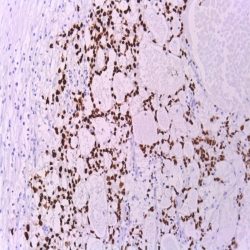
آنتی بادی S100-Beta Protein (SP127)
Name:Antibody S100-Beta Protein clone SP127
Description and applications: S100 proteins are a set of 25 small acidic molecules with a molecular mass of 10-12 kDa that are functionally grouped into homo or heterodimers composed of two subunits, alpha and beta, capable of combining with each other and with extensive sequence homology. There exist up to 14 variants of the alpha chain gene, which is located on chromosome 1q21; on the contrary, there is only one sequence of the beta chain gene, located on chromosome 21q22.3, showing its greater functional specificity. In its dimeric form, the S100 protein, together with calmodulin and troponin C, belongs to the calcium binding protein family; its affinity for this ion, as well as for other metals such as zinc, is remarkable. It is for this reason that the S100 protein is involved in basic cellular activities such as cation diffusion across lipid membranes, microtubule assembly, and RNA polymerase activity. In neurons, the S100 protein also regulates the interaction between chromosomes and synaptosomes. Depending on the combination between the alpha and beta single chains to form dimers and the homology of their sequences, there exist three forms of S-100 protein: A, with at least 15 subtypes between A1 and A14, B, and G. The S100-B protein is a 21-kDa beta-chain homodimer involved in the regulation of numerous processes that require calcium ion flow stimulation. The S100-B protein is present in neurons, glial cells of the central nervous system, Schwann cells and satellite cells of the peripheral nervous system (but not perineural cells), melanocytes, myoepithelial cells, glandular epithelia of the breast and the kidney, skeletal muscle and heart cells, adipocytes, and chondrocytes. This molecule is also expressed in antigen-presenting cells such as Langerhans cells of the skin, and interdigitating dendritic cells in the T-cell areas of lymphoid tissue. In contrast, the S100-B protein is not present in smooth muscle fibres. In neoplasms, the S100-B protein is highly expressed in tumours of the central and peripheral nervous systems and in melanomas, including desmoplastic melanoma. However, up to 4% of melanomas may be negative against this antibody, suggesting the need to assess this staining within a minimal panel of antimelanoma antibodies that at least includes the HMB45 and Melan A markers. Likewise and in comparison with primary lesions, some loss of expression of the S100-B protein has been observed in metastatic melanoma lesions. The S100-B protein is also useful in the diagnosis of tumours derived from cartilage, adipose tissue, and interdigitating reticular cells in lymphoid tissue, as well as granular cell peripheral nerve sheath tumours, myoepithelial tumours, paragangliomas (sustentacular cells), PEComas, Rosai-Dorfman disease, clear cell sarcoma, chordoma, adenoid cystic carcinoma, and isolated cases of synovial sarcoma and rhabdomyosarcoma. Atypical fibroxanthomas, cardiac sarcomas, myofibroblastomas, xanthomas, and solitary fibrous tumours are all negative for S100-B. Finally, altered overexpression of the beta-chain gene with S100-B protein overexpression may be found in the basis of some of the lesions observed in various types of neurological and genetic diseases such as Alzheimer’s disease, Down’s syndrome (trisomy 21), epilepsy, amyotrophic lateral sclerosis, and type I diabetes, in which an excess of the S100-B protein has a potential neurotoxic effect. In this sense, several tests have been developed for serum determination of this cell damage marker in these patients.
Composition: Anti-human S100-Beta Protein rabbit monoclonal antibody purified from serum and prepared in 10mM PBS, pH 7.4, with 0.2% BSA and 0.09% sodium azide
-
 آنتی بادیهای ایمونوهیستوشیمی
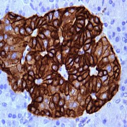
آنتی بادی S100-P Protein (16/F5)
Name: Antibody Protein S100 clone 16/F5
Composition: Anti-human S100-P Protein mouse monoclonal antibody purified from serum and prepared in 10mM PBS, pH 7.4, with 0.2% BSA and 0.09% sodium azide
Description and applications: The S100P protein, or placental S100, which is a member of the large S100 protein family, is involved in Ca2+ intracellular transport and, together with EZR and PPP5C proteins, participates in the formation of microvilli in epithelial cells and stimulates apocrine cell secretion. Unlike the genes of the other members of the S100 family of proteins, which co-localise in the 1q21 region, the S100P protein is encoded by the S100P gene, located in the 4p16 region. The 100 family of proteins is expressed in a wide range of cells, where it plays a role in cell cycle progression and in cell differentiation. The S100P molecule was initially identified in the human placenta at rather high levels. Pathologically, the antibody against the S100P protein has shown nuclear or nuclear/cytoplasmic immunoreactivity in almost all pancreatic ductal adenocarcinomas, while it displays no staining in normal ducts and in pancreatic acini. In addition, the S100P protein has also been detected in virtually all pancreatic intraductal papillary mucinous neoplasms, as well as in its microinvasive and diffusely invasive component, including tumours with perineural and lymphatic invasion. For both reasons, the antibody against the S100P protein is considered a useful tool for the differential diagnosis between reactive and neoplastic lesions of the pancreas. In the case of liver and bile ducts, the antibody also exhibits marked nuclear and cytoplasmic positivity in malignant lesions derived from bile ducts, while the benign epithelium derived from these tumours is negative. Similarly, detection of both S100P and GATA3 may help to distinguish urothelial carcinomas from other genitourinary neoplasms such as prostate cancers and renal cell carcinomas; in this case, however, and in contrast to what is observed in biliary and pancreatic ductal lesions, S100P expression can be detected in the normal urothelium. Finally, the anti-S100P antibody can be used as a prognostic factor in non-small cell lung carcinomas, where S100P expression has been associated with frequent distant metastases and short survival.
-
 آنتی بادیهای ایمونوهیستوشیمی
آنتی بادی SALL4 (EE-30)
Name: Antibody SALL4 clone EE-30
Description and aplications: Sall3 (SALL3, sal-like 3) and Sall4 (SALL4, sal-like 4) are mammalian homologs of the Drosophila region-specific homeotic gene spalt, which encodes a zinc fingercontaining transcription regulator. Drosophila spalt is an essential genetic component required for the specification of posterior head and anterior tail as opposed to trunk. Sall3 is expressed at 24 weeks of gestation in several regions of the human fetal brain including neurons of the hippocampus formation and of mediodorsal and ventrolateral thalamic nuclei, Purkinje cells of the cerebellum and a subset of neurons in the brainstem. Sall4 expression in early mouse embryos is gradually confined to the head region and the primitive streak, followed by prominent expression in the developing midbrain, branchial arches, limbs and genital papilla. SALL4 has been considered as a pan-marker for germ cell tumors. However, positivity might occur in undifferentiated neoplasm of digestive or urogenital system.
Composition: anti-human SALL4 mouse monoclonal antibody prepared in 10mM PBS, pH 7.4, with 0.2% BSA and 0.09% sodium azide
-
 آنتی بادیهای ایمونوهیستوشیمی
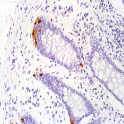
آنتی بادی Serotonin (5HT-H209)
Name:Antibody Serotonin clone 5HT-H209
Description and applications: Serotonin-positive cells are present in gastric, duodenal, jejunal, ileal, colonic, and appendiceal mucosae. Positive cells are sparse and usually occur in the lower half of the gastric and intestinal mucosae. In the appendix, in addition to the mucosal serotonincontaining cells, there are single or small clusters of serotoninpositive cells in the sub-epithelial region of the lamina propria.
Composition: Anti-human Serotonin mouse monoclonal antibody purified from serum and prepared in 10mM PBS, pH 7.4, with 0.2% BSA and 0.09% sodium azide
-
 آنتی بادیهای ایمونوهیستوشیمی
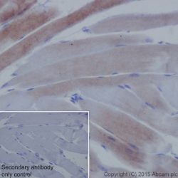
آنتی بادی Skeletal Muscle Actin (5C5,F8,C7)
Name:Antibody Skeletal Muscle Actin clone 5C5,F8,C7
Description and applications: 5C5.F8.C7 MAb is highly specific and shows no crossreaction with smooth muscle actin. This antibody reacts with sarcomeric actins of normal tissues and neoplasms derived from such tissues (i.e. rhabdomyosarcomas).
Composition: Anti-human Skeletal Muscle Actin mouse monoclonal antibody purified from serum and prepared in 10mM PBS, pH 7.4, with 0.2% BSA and 0.09% sodium azide
-
 آنتی بادیهای ایمونوهیستوشیمی
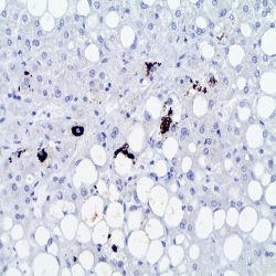
آنتی بادی P62 (3/P62 LCK LIGAND)
Name: P62 Antibody clone LCK LIGAND 3/P62
Description and applications: This antibody recognises phosphotyrosine p62, a member of the c-src family of cytoplasmic kinases, which binds as a ligand to the SH2 domains of p56 (Lcr) in the absence of phosphotyrosine. p62/SQSTM1 is an adapter protein targeting the protein aggregates ubiquitinated by lysosomal degradation. p62/SQSTM1 is selectively degraded via the autophagic pathway. Mallory’s hyaline and intracellular hyaline bodies are associated with chronic non-neoplastic liver diseases (steatohepatitis, related or not to alcohol abuse; toxic or metabolic chronic cholestasis) and with neoplastic liver diseases such as hepatocellular carcinoma. These inclusions are related to and contain keratin aggregates, especially keratin 8, ubiquitin, heat shock proteins, and the stress adaptor protein p62, stabilized by bonds catalysed by transglutaminases. P62 binds to ubiquitin and acts as an adaptor to bind ubiquitinated proteins. Autophagy is an evolutionary highly conserved degradative mechanism affecting the components of the cytoplasm that contributes to homoeostasis by making organelle replacement possible. Unlike the ubiquitin-proteosome system, the autophagy mechanism occurs in multiple steps that include the formation of the phagophore, which traps proteins and organelles for its degradation, and the autophagosome, which, through mechanisms involving cytoskeleton’s microtubules, fuses with lysosomes to form autolysosomes, where the material is eventually degraded. This antibody is useful for the identification of p62, found in Mallory’s hyaline and intracellular hyaline bodies present in chronic liver diseases and hepatocellular carcinomas. It is also useful to detect p62 in autophagic vacuoles present in various neurodegenerative, neoplastic infectious, and inflammatory neuromuscular diseases.
Composition: Anti-human P62 mouse monoclonal antibody purified from serum and prepared in 10mM PBS, pH 7.4, with 0.2% BSA and 0.09% sodium azide
-
 آنتی بادیهای ایمونوهیستوشیمی
آنتی بادی p63 (4A4)
Name: Mouse anti-human p63 Monoclonal Antibody clone 4A4
Description and aplications: p63, a homolog of the tumor suppressor p53, has been identified in basal cells in the epithelial layers of a variety of tissues, including epidermis, cervix, urothelium, breast and prostate. p63 was detected in nuclei of the basal epithelium in normal prostate glands; however, it was not expressed in malignant tumors of the prostate. As a result, p63 has been reported as a useful marker for differentiating benign from malignant lesions in the prostate, particularly when used in combination with markers of high molecular weight cytokeratins and the prostate-specific marker AMACR (P504S). p63 has also been shown to be a sensitive marker for lung squamous cell carcinomas (SqCC), with reported sensitivities of 80-100%. Specificity for lung SqCC, vs. lung adenocarcinoma, has been reported to be approximately 70-90%, as positive staining with p63 has been typically observed in 10-30% of lung adenocarcinomas cases. In breast tissue, p63 has been identified in myoepithelial cells of normal ducts. Reports have described the utility of p63 in a panel of IHC markers for the assessment of breast lesions, due to the differential expression of the luminal vs. basal and myoepithelial markers.
Composition: anti-human p63 mouse monoclonal antibody purified from ascites. Prepared in 10mM PBS, pH 7.4, with 0.2% BSA and 0.09% sodium azide
-
 آنتی بادیهای ایمونوهیستوشیمی
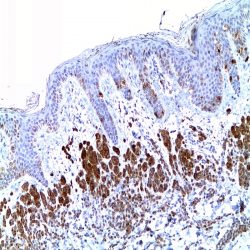
آنتی بادی Pan Melanoma Cocktail (HMB45+M2-7C10+M2-9E3)
Name: Pan Melanoma Cocktail Antibody clone HMB45+M2-7C10+M2-9E3
Description and applications: The antibody represents a cocktail of anti-gp100 (HMB45) and MART-1 antibodies. By
immunohistochemistry, it recognizes melanocytes and melanomas. This cocktail reacts with junctional and blue nevus cells and with fetal and neonatal melanocytes. It does not stain tumor cells of epithelial, lymphoid, glial, or mesenchymal origin. The antibody labels melanotic as well as amelanotic variants of melanomas. It is extremely sensitive for labeling formalin-fixed, paraffinembedded melanomas and other tumors showing melanocytic differentiation.Composition: Anti-human Pan Melanoma Cocktail mouse monoclonal antibody purified from serum and prepared in 10mM PBS, pH 7.4, with 0.2% BSA and 0.09% sodium azide
-
 آنتی بادیهای ایمونوهیستوشیمی
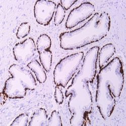
آنتیبادی Pan-Cadherin (CH-19)
Name:Pan-cadherin Antibody clone CH-19
Description and applications: Cadherins are a broad family of cell-cell adhesion molecules with varying degrees of tissue expression. E-cadherin is found in all normal epithelia, whereas Pcadherin is expressed in trophoblastic cells and shows limited expression in epithelia, being predominantly expressed in bronchial and mammary basal epithelia. Cadherins are calcium-dependent adhesion molecules; intracellularly, E-cadherin binds to members of the catenin family (β, γ or α-catenins), which in turn are connected to the cytoskeleton’s actin filaments. Cadherins play an important role in cell growth and development, maintaining tissue integrity and architecture. Loss of cadherin expression plays an important role in neoplasms, facilitating local invasion and metastases generation. Immunohistochemical studies on carcinomas show decreased cadherin expression compared to normal tissues, especially in highly invasive tumours such as gastric adenocarcinomas.
Composition: Anti-human Pan-Cadherin mouse monoclonal antibody purified from serum and prepared in 10mM PBS, pH 7.4, with 0.2% BSA and 0.09% sodium azide
-
 آنتی بادیهای ایمونوهیستوشیمی
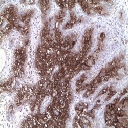
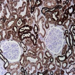
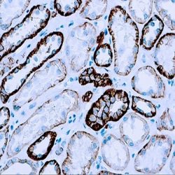
آنتی بادی Pancreatic lipase (EPR6275)
Name: Pancreatic lipase Antibody clone EPR6275
Description and applications: Pancreatic lipase is a 56kDa molecular mass enzyme actively secreted in the duodenum by the pancreas in the presence of bile salts and colipase. It belongs to the family of triglyceride lipases, of which there are at least three tissue-specific isoenzymes (pancreatic, hepatic and gastric and lingual) that break down dietary fats by converting triglycerides into monoglycerides and free fatty acids. Pancreatic lipase together with bile salts favors the absorption into the lymphatic system of the monomers resulting from digestion. Human pancreatic lipase is encoded by the PNLIP gene of 13 exons and more than 20 kb, located on the chromosomal region 10q25.3. In normal tissues this antibody identifies pancreatic acinar cells and in neoplasms it recognizes acinary cell carcinoma of the pancreas, which expresses this marker in more than 50% of cases. For this reason and integrated in a panel of suitable antibodies, pancreatic lipase allows the differentiation of acinar adenocarcinoma of pancreas from other malignancies with similar morphology (especially neuroendocrine carcinoma with acinary architecture) and establish the pancreatic origin of distant metastases, that on numerous occasions may be the first manifestation of the tumor.
Composition: Anti-human Pancreatic lipase mouse monoclonal antibody purified from serum and prepared in 10mM PBS, pH 7.4, with 0.2% BSA and 0.09% sodium azide
-
 آنتی بادیهای ایمونوهیستوشیمی
آنتی بادی Pancreatic Polypeptide (Polyclonal)
Name:Pancreatic Polypeptide Antibody
Description and applications: This gene belongs to the NPY family and it encodes a protein that is synthesized as a 95 aa polypeptide precursor in the pancreatic islets of Langerhans. It is cleaved into two peptide products; the active hormone of 36 aa and an icosapeptide of unknown function. The hormone acts as a regulator of pancreatic and gastrointestinal functions and may be important in the regulation of food intake. Plasma level of this hormone has been shown to be reduced in conditions associated with increased food intake and elevated in anorexia nervosa. In addition, infusion of this hormone in obese rodents has shown to decrease weight gain.
Composition: anti-human Pancreatic Polypeptide goat polyclonal antibody purified from serum and prepared in 10mM PBS, pH 7.4, with 0.2% BSA and 0.09% sodium azide
Immunogen: Synthetic peptide sequence (TRPRYGKRHKEDT) corresponding to the internal amino acids of PPY
-
 آنتی بادیهای ایمونوهیستوشیمی
آنتی بادی Cytokeratin 14 (LL002)
Name: Cytokeratin 14 antibody clone LL002
Description and aplications: Cytokeratin 14 belongs to the type A (acidic) subfamily of low molecular weight keratins and exists in combination with keratin 5. Cytokeratin 14 has been studied as a prognostic marker in breast cancer. Here it is used as a marker for the myoepithelial cells and for the identification of basaloid histotype of breast carcinoma.
The antibody also distinguishes stratified epithelial cells from simple epithelial cells and has been reported useful in the identification of squamous cell carcinomas.Composition: anti-human Cytokeratin 14 mouse monoclonal antibody purified from ascites fluid by Protein G chromatography. Prepared in 10mM PBS, pH 7.4, with 0.2% BSA and 0.09% sodium azide
Immunogen: A synthetic peptide of 15 amino acid residues from the C-terminus of human keratin 14.
-
 آنتی بادیهای ایمونوهیستوشیمی
آنتی بادی Cytokeratin 5/6/8/18 (EP24/EP67/B22.1&B23.1)
Name:Cytokeratin Antibody clone EP24/EP67/B22.1&B23.1
Description and applications: This antibody recognises cytokeratins 5, 6, 8, and 18. Proteins generically called intermediate filaments, because they measure between 7 and 22 nm in diameter (i.e., with a size between actin, 5-7 nm, and tubulin, 22-25 nm), are part, along with actin and tubulin, of the vertebrate cytoskeleton. This superfamily is composed of six subfamilies of molecules with different tissue expression patterns. Cytokeratins constitute homology groups I and II, and in humans they are encoded by more than 49 different genes located in chromosomes 17 (I) and 12 (II). The nomenclature chosen in 1982 by Moll and Franke assigns numbers from 1 to 8 to type II cytokeratins (neutral or basic) and from 9 to 21 to type I cytokeratins (acidic). An analogous nomenclature for hair keratins has now been defined, with the addition of letters Ha and Hb to distinguish type I from type II. Structurally, cytokeratins share with the rest of intermediate filaments a central axis of 310 amino acid residues consisting of four highly preserved α-helical domains (1A, 1B, 2A, and 2B) that define the type of intermediate filament that will be formed after assembly; these domains are separated by three non-helical linking regions (L1, L12, and L2) and two end domains which are highly different in size and sequence (head, 1, and tail, 2), each with constant (E1/E2), variable (V1/V2), and homology (H1/H2) regions, the latter characteristic of type II keratins and absent in type I keratins. The major immunogenic properties and the main differences between each keratin class are found in the variable domains. Keratins are usually assembled into I/II heterodimers and are specifically co-expressed in pairs in each tissue. Due to the high homology among the different molecules, it is common for a monoclonal antibody to react with different types of cytokeratins; for example, anti-cytokeratin AE1 labels cytokeratins 10, 14, 15, 16, and 19. The anti-cytokeratin monoclonal antibody cocktail is a broad-spectrum antibodt for general use. This antibody mixture reacts with carcinomas in general, but as with other anti-cytokeratin antibodies, it is possible that poorly differentiated carcinomas occasionally exhibit poor immunostaining. Its most common application is the differential diagnosis of malignant tumours, in which case it can be used within a panel of antibodies, such as anti-ALC, which recognises lymphoma, or anti-S100 or HMB45, which labels malignant melanoma. The staining of other non-carcinoma tumours is uncommon, although as with other anti-cytokeratin 8 antibodies, smooth muscle and leiomyoma labelling may occasionally be observed.
Composition: Anti-human Cytokeratin 5/6/8/18 rabbit/mouse monoclonal antibody purified from serum and prepared in 10mM PBS, pH 7.4, with 0.2% BSA and 0.09% sodium azide
-
 آنتی بادیهای ایمونوهیستوشیمی
آنتی بادی ACTH (Adrenocorticotropic Hormone) (Polyclonal)
Name: Rabbit anti-human ACTH (Adrenocorticotrophic Hormone) Polyclonal Antibody
Description and applications: Anti-ACTH is a useful marker in classification of pituitary tumors and the study of pituitary disease. It reacts with ACTH-producing cells (corticotrophs). It also may react with other tumors (e.g., some small cell carcinomas of the lung) causing paraneoplastic syndromes by secreting ACTH.
Composition: anti-human ACTH rabbit monoclonal antibody purified from serum and prepared in 10mM PBS, pH 7.4, with 0.2% BSA and 0.09% sodium azide
Intended use : Immunohistochemistry (IHC) on paraffin embedded tissues. Not tested on frozen tissues or Western-Blotting
Immunogen: Synthetic peptide derived from Nterminus of human ACTH.
-
 آنتی بادیهای ایمونوهیستوشیمی
آنتی بادی Calponin (EP63)
Name:Antibody Calponin clone EP63
Description and applications: Calponin is a smooth muscle specific, actin-, tropomyosin- and calmodulin-binding protein thought to be involved in regulation of actomyosin as well as the regulation or modulation of contraction. It is expressed on smooth muscle cells and myoepithelial cells. Calponin antibody has been used to identify invasion of breast lesion. Additionally, Calponin is expressed on malignant fibrous histiocytoma of bone and adenoid cystic carcinoma of salivary gland. The consistently positive staining pattern in adenoid cystic carcinomas may be useful in discriminating
Composition: Anti-human Calponin rabbit monoclonal antibody purified from serum and prepared in 10mM PBS, pH 7.4, with 0.2% BSA and 0.09% sodium azide
-
 آنتی بادیهای ایمونوهیستوشیمی
آنتی بادی Delta Catenin-1 (p120) (EP66)
Name: Delta Catenin-1 (p120) (Clone EP66)
Description and applications:Catenins are proteins that are linked to the cytoplasmic domain of transmembrane cadherins. p120 Catenin is a member of this Armadillo gene family of junctional plaque proteins. The association of catenins to cadherins produces a complex which is linked to the actin filament network, and which seems to be important for cadherins cell-adhesion properties. Cytoplasmic accumulation of p120 Catenin has been observed in lung cancer, pancreatic cancer, gastric cancer and colon cancers and is associated with poor progress in colon cancer patients. In breast lobular neoplasia, anti p120 Catenin shows a diffuse cytoplasmic immunostaining pattern, while breast ductal neoplasia retains the membrane immunostaining pattern. p120 Catenin antibody is useful in differentiation of lobular carcinoma from ductal carcinoma of the breast and in identifying early lesions of lobular neoplasia.
Composition:Anti-human Delta Catenin-1 (p120) rabbit monoclonal antibody purified from serum and prepared in 10mM PBS, pH 7.4, with 0.2% BSA and 0.09% sodium azide.
Immunogen: A synthetic peptide corresponding to residues in human p120 Catenin protein.
-
 آنتی بادیهای ایمونوهیستوشیمی
آنتی بادی Epithelial Membrane Antigen (EMA) (E29)
Name: Epithelial Membrane Antigen(EMA)-CloneE29
Description and applications: Anti-EMA antibody is a useful marker for staining many carcinomas. It stains normal and neoplastic cells from various tissues, including mammary epithelium, sweat glands and squamous epithelium. Hepatocellular carcinoma, adrenal carcinoma and embryonal carcinomas are consistently EMA negative, so keratin positivity with negative EMA favours one of these tumours. EMA is frequently positive in meningioma, which can be useful when distinguishing it from other intracranial neoplasms, e.g. Schwannomas. The absence of EMA can also be of value since negative EMA staining is characteristic of some tumours including adrenal carcinoma, seminomas, paraganglioma and hepatoma. Many mesotheliomas, epithelioid and synovial sarcomas, chordomas, choroid plexus tumors, nodular lymphocyte-predominant Hodgkin lymphoma, anaplastic CD30 / ALK positive lymphomas and myelomas are positive; occasionally some small round cell tumours and small cell sarcomas, T null phenotype or CD56 / CD57 + T / NK lymphomas are positive. Other lymphomas, basal cell carcinomas, hepatocellular carcinomas, melanomas, endocrine neoplasms and soft tissue tumours are consistently negative.
Composition: anti-human EMA mouse monoclonal antibody purified from serum and prepared in 10mM PBS, pH 7.4, with 0.2% BSA and 0.09% sodium azide.
برای تماس کلیک کنید
سما تشخیص آریا ، عرضه کننده متنوع ترین و کامل ترین محصولات کیت های آزمایشگاهی