برای تماس کلیک کنید

Welcome to Sama Tashkhis Aria Official Website

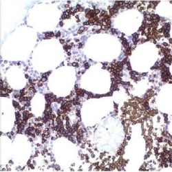
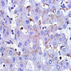
آنتی­ بادی MUM1/IRF4 (MUM1p)

Name: MUM1 Antibody (Clone MUM1p)

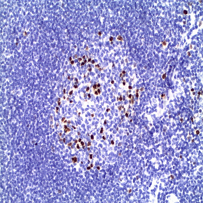
Description and aplications: MUM1 / IRF4 is an oncogene associated with myeloma. The protein has a molecular weight of 51.6 kDa and is encoded by a gene on chromosome 6p25-p23 region located mainly in the nucleus of lymphocytes. When MUM1 is not expressed, the B / T cells do not get activated and the immunoglobulins are not secreted by the plasma cells. Most neoplasms composed of mature lymphoid cells express MUM1. Therefore, this protein is expressed in a subset of B cells in the germinal center in plasma cells and in activated T cells. MUM1 is a marker that helps in characterizing the histogenesis of lymphoma / leukemia of B origin. In Bcell lymphomas, MUM1 expression presents a predictive role of the genetic profile and represents, together with CD10 and BCL-6, the base of Hans classification for diffused large B cell lymphomas. MUM1 positivity, in these cases, suggests a non-germinal center phenotype and is predictive for prognosis, especially in pediatric cases. In Burkitt lymphomas, that shares cytological, architectural and histological aspects with some cases of DLBCL of “Burkitt-like”, the MUM1 is negative in general or positive in a small percentage of cases or tumoral cells. All cases of primary effusion lymphomas and most cases of primary brain lymphomas are MUM1 positive. Other lymphoid neoplasm express MUM1. Melanomas can also stain with MUM1.

Composition: anti-MUM1 mouse monoclonal antibody obtained from supernatant culture and prediluted in a tris buffered solution pH 7.4 containing 0.375mM sodium azide solution as bacteriostatic and bactericidal.

حجم موجود3 / 7 / 12 میلی‌لیتر

مورد استفادهایمونوهیستوشیمی روی بافت پارافین. بر روی بافتهای فروزن یا وسترن بلات تست نشده است.

کلون: MUM1p

ایزوتایپ: Mouse IgG1kappa

کنترل مثبت IHC: تونسیل

قابلیت دیده شدن: هسته ای

روش پیشنهادی IHC:

  • برش تهیه شده با ضخامت μm4 باید روی لام شارژ شده در طول شب و در دمای 60 درجه سانتی گراد خشک شود.
  • دپارافینه کردن، رهیدراته کردن و استفاده از حرارت جهت بازیابی اپی توپ : بافت را با استفاده از بافر EDTA pH 8 برای بیست دقیقه در دمای 95 درجه سانتی گراد بجوشانید. سپس 5-3 بار با استفاده از آب مقطر یا دیونیزه شده شسته و سپس بیست دقیقه در دمای اتاق خنک کنید.
  • بلاک پروکسیدازهای درونی : بلاک با استفاده از محلول پروکسیداز به مدت ده دقیقه
  • آنتی بادی اولیه: برای 10 دقیقه در انکوبه کنید پروتکل ممکن است بسته به تهیه نمونه و کاربرد خاص متفاوت باشد. شرایط استاندارد باتوجه به آزمایشگاه و کاربر متفاوت می باشد.
  • برای رنگ آمیزی ، می توانید از کیت Master Polymer Plus Detection یا کیتهای دیتکشن سایر کمپانی ها استفاده کنید.
  • رنگ میزی نهایی با هماتوکسیلین و فیکس نهایی اسلاید.

شرایط نگهداری و پایداری : حداکثر 18 ماه؛ در دمای 2-8 درجه سانتیگراد نگهداری شود. فریز نشود.


این آنتی‌بادی متعلق به پنل‌‌‌‌‌‌ تشخیصی LYMPHOID است.‌


Download Cataloge

Icon

MUM1/IRF4 (MUM1p) 565.77 KB 4 downloads

...
.well.c2a3 .btn { padding: 11px 30px; font-size: 11pt; } .well.c2a3 .media-body { font-size: 11pt; } .well.c2a3 .wpdm_icon { height: 42px; width: auto; }

Reviews

There are no reviews yet.

Be the first to review “آنتی­ بادی MUM1/IRF4 (MUM1p)”

Your email address will not be published. Required fields are marked *

Scroll to Top