-
 آنتی بادیهای ایمونوهیستوشیمی
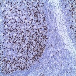
آنتی بادی CD21 (2G9)
Name: CD21 Antibody Clone 2G9
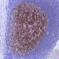
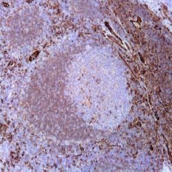
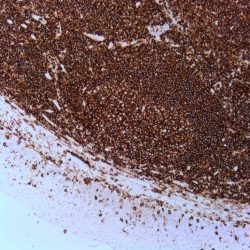
Description and applications: The CD21 or CR2 antigen is a 145 kD glycoprotein of molecular weight, which constitutes one of the
membrane molecules involved in the transduction of growth signals into the B cells. Also acts as a receptor for the Epstein Barr virus. This antibody also reacts with a leukocyte membrane epitope (CR2), with molecular weights of 95, 72, 50, 32 and 28 kD. The 28 and 72 kD molecular weight fragments of CR2 constitute the point of attachment of C3d. CD21 antigen is an antigen that is expressed in mature B cells. The antigen is present with a high density in follicular dendritic cells (CFD) of B zones of lymphoid tissue. CD21 moderately stains B cells and strongly follicular dendritic cells in frozen tissue sections, while staining of B cells is reduced or non existent in the paraffin embedded sections. Immunostaining of CFDsin paraffin-embedded sections is as strong as that of frozen tissue sections. CD21 immunohistological analysis of CFD of non- Hodgkin’s lymphomas in paraffin embedded sections reveals the generally dense and highly defined nodular network of CFD in follicular lymphomas and a diffuse, disorganised network in some types of diffuse lymphomas. B-cell precursor lymphomas (lymphoblastic lymphomas), Burkitt’s lymphomas, plasmacytomas, and hairy cell leukemias are
constantly lacking a CFD. In non-Hodgkin’s lymphomas of T cell origin, CFDs are mainly restricted to peripheral T-cell lymphomas of the
angioinmunoblastic type and some cases of pleomorphic lymphomas. T-cell lymphoblastic lymphomas, T-CLL and mycosis fungoides do not have a CFD network. The only category of Hodgkin’s disease that has a CFD-like network is the nodular lymphocyte-predominant
form.Composition: Anti-human CD21 mouse monoclonal antibody purified from serum and prepared in 10mM PBS, pH 7.4, with 0.2% BSA and 0.09% sodium azide.
Immunogen: Recombinant human CD21.
-
 آنتی بادیهای ایمونوهیستوشیمی
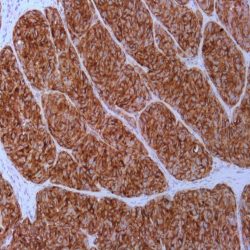
آنتی بادی CD27 (EPR8569)
Name: CD27 Antibody Clone EPR8569
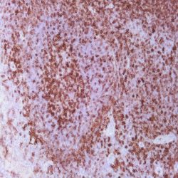
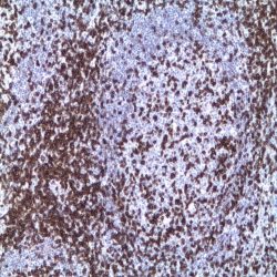
Description and applications: CD27 is a glycosylated, type I transmembrane protein with a molecular mass of 50-55 kDa that belongs to the TNF receptor superfamily (TNFRSF7), of which it is its seventh member. This protein is also known as T cell activation antigen, S152 protein and T14 protein. It is encoded by a gene located in the chromosomal region 12p13.31. As a member of the tumour necrosis factor (TNF) receptor family, CD27 plays an important role in cell growth and differentiation as well as in apoptosis regulation. In the former case, CD27 binds to CD70, its ligand, and plays an important role in costimulation of T cell activation and regulation of B cell differentiation and proliferation and immunoglobulin synthesis. The cytoplasmic domains of CD27 have also been shown to interact with TRAF2 and TRAF5 to promote activation of NF-kappa B and SAPK/JNK. Its role inapoptosis is linked to its ability to bind to the proapoptotic SIVA protein; thus, CD27 plays an important role in inducing this cell death process. CD27 is expressed in medullary thymocytes, virtually all mature T cells, memory B cells, NK cells, and a fraction of normal plasma cells but not in virgin B cells or effector T cells. In patients diagnosed with acute lymphoblastic leukaemia (ALL), overexpression of CD27 is associated with a more favourable prognosis and an increased anti-leukaemic immune response by the organism. Immunohistochemical techniques have confirmed the expression of the CD27 antigen in isolated cases of mantle cell lymphoma, Burkitt lymphoma, marginal zone lymphoma, and plasmacytoma. In cases of multiple myeloma, apparently the expression of CD27 has predictive value for patient survival, with an overall three-year survival rate of 92% in positive cases compared to 50% in negative cases. CD27 negativity in cases of hairy-cell leukaemia diagnosed in the spleen has been suggested as a useful tool in the differential diagnosis with other types of B cell leukaemia.
Composition: Anti-human CD27 mouse monoclonal antibody purified from serum and prepared in 10mM PBS, pH 7.4, with 0.2% BSA and 0.09% sodium azide.
Immunogen:Human recombinant protein similar to CD27.
-
 آنتی بادیهای ایمونوهیستوشیمی
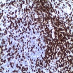
آنتی بادی CD278 (SP98)
Name: CD278 Antibody Clone SP98
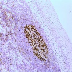
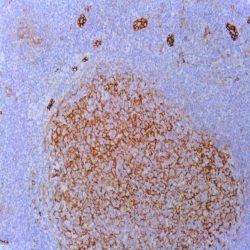
Description and applications: CD278 belongs to the CD28 and CTLA-4 cell-surface receptor family and is highly expressed in activated T cells. It is essential both for efficient interaction between T and B cells and for normal antibody responses to T cell dependent antigens. It enhances all basic T cell responses to a foreign antigen, namely proliferation, secretion of lymphokines, up-regulation of molecules that mediate cell-cell interaction, and effective help for antibody secretion by B cells.
Composition: Anti-human CD278 rabbit monoclonal antibody purified from serum and prepared in 10mM PBS, pH 7.4, with 0.2% BSA and 0.09% sodium azide.
Immunogen: Synthetic peptide corresponding to the C-terminus of human CD278.
-
 آنتی بادیهای ایمونوهیستوشیمی
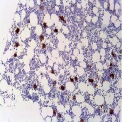
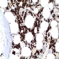
آنتی بادی CD30 (Ber-H2)
Name: CD30 Antibody Clone Ber-H2
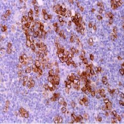
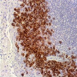
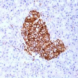
Description and aplications: The CD30 antigen (also called Ki-1) is a single chain glycoprotein that belongs to the family of for tumor necrosis receptors factors (TNF)
and acts as a type 1 transmembrane receptor. The gene encoding CD30 has been mapped to chromosome 1p36. The CD30 glycoprotein is synthesized as a 90 kDa precursor that is transformed into the Golgi complex in a mature 105/120 kDa phosphorylated glycoprotein that binds to the cell membrane. In normal tissues CD30 expression is restricted to some immunoblasts (both B and T) located around lymphoid follicles of antigenically stimulated lymphoid tissue, acinar cells of the pancreas and during pregnancy, by some decidual cells. In neoplasms, the CD30 antigen is expressed in anaplastic tumors, such as the mono- bi- and multinucleated cells of Hodgkin’s disease cells and thevast majority of anaplastic large cell lymphoma, a third of which lack the CD45 immunostaining. The CD30 antigen is also expressed in some non-hematopoietic malignancies such as embryonal carcinomas and rarely in poorly differentiated tumors of epithelial origin or melanocytes. For this reason and for in vitro diagnosis, the CD30 antibody should be used as part of an ample panel of antibodies that rule out the possibility of nonlymphoid origin for positive tumors.Composition: anti-human CD30 mouse monoclonal antibody purified from ascites fluid by Protein A chromatography. Prepared in 10mM PBS, pH 7.4, with
0.2% BSA and 0.09% sodium azide.Immunogen: Co cells.
-
 آنتی بادیهای ایمونوهیستوشیمی
آنتی بادی CD31/PECAM-1 (JC/70A)
Name: CD31 Antibody Clone JC/70A
Description and aplications: CD31 is a glycoprotein expressed on endothelial cells and in platelets. It is known to be involved in cell signaling and cell adhesion. The anti-CD31 antibody is useful for the identification of benign and malignant vascular tumors. Other lesions like histiocytosis X, sinus histiocytosis with massive lymphadenopathy or Rosai-Dorfman disease, plasmacytoma and multiple myeloma are also frequently positive to CD31.
Composition: anti-human CD31 mouse monoclonal antibody purified from culture supernatant. Prepared in 10mM PBS, pH 7.4, with 0.2% BSA and 0.09% sodium azide.
Immunogen: Membrane preparation of a spleen from a patient with hairy cell leukemia.
-
 آنتی بادیهای ایمونوهیستوشیمی
آنتی بادی CD34 (QB-End/10)
Name:CD34 Antibody clone QBEnd/10
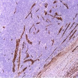
Description and aplications: The QBEnd10 antibody is a class II monoclonal antibody that recognizes an epitope of CD34 that is resistant to neuraminidase and sensitive to glycoprotease and chymopapain. CD34, a single chain transmembrane glycoprotein, is detected in bone marrow precursors, including normal lymphoid precursors or hematogonias where it may act as lectins ligand of these cells with the bone marrow stroma. CD34 expression disappears from the surface of all haematological series during maturation. Megakaryocytes may show varying positivity for CD34. This antibody shows reactivity with most endothelial
cells, expressed on the luminal surface and interdigitating membrane processes between endothelial cells, but is absent in large venous and arterial vessels. CD34 is also absent in sinusoids of the spleen, liver and placenta. The lymphatic vessels are generally weakly stained. CD34 is also expressed in fibroblast-like dendritic cells localized in liver portal tracts and in Peyer’s patches in wound repair phase. In tumors, CD34 is detected in myeloid blasts of myelodysplastic syndromes and in most cases of acute myeloid leukemias and lymphoblast of the acute lymphoblastic leukemias. Mature B cell lymphomas, B and T cell leukemias are all negative for CD34.
Most vascular tumors and sarcomas including Kaposi hemangiosarcomas are positive for CD34. CD34 is expressed in most cases of dermatofibrosarcoma protuberans (while fibrous histiocytoma is negative), solitary fibrous tumor, lipomas (particularly the spindle cell) and liposarcomas, gastrointestinal stromal (GIST) (strongly positive in 80% of cases coinciding with the expression of CD117) and meningiomas in a variable proportion of cases. Moderate expression was detected in cutaneous leiomyomas and weak staining may be observed in tumors of smooth muscle of the uterus or soft tissue.Composition: anti-human CD34 mouse monoclonal antibody purified from ascites fluid by Protein A chromatography. Prepared in 10mM PBS, pH 7.4, with
0.2% BSA and 0.09% sodium azideImmunogen: Detergent solubilized vesicular suspension prepared from a perfusate of human term placenta.
-
 آنتی بادیهای ایمونوهیستوشیمی
آنتی بادی CD38 (38C03)
Name: CD38 Antibody Clone 38C03(same as SPC32)
Description and applications: CD38 is a single chain type II integral transmembrane protein, which is highly expressed on thymocytes. It is also present on activated T cells and terminally differentiated B cells (plasma cells). Other reactive cells include NK cells, monocytes, macrophages, and dendritic cells. CD38 may be detected on cells from multiple myeloma, ALL (B and T) and some AML. It is, however, not found on most mature resting peripheral lymphocytes. CD38 functions as a multicatalytic ectoenzyme serving as ADP-ribosyl cyclase, cyclic ADP-ribose hydrolase and possibly NAD+ glycohydrolase or as a cell surface receptor.
Composition: Mouse anti-human CD38 monoclonal antibody is obtained from tissue culture supernatant purified and diluted in 10 mM Phosphate buffered saline (PBS), pH 7.2 containing 1% bovine serum albumin (BSA) and 0.09% sodium azide (NaN3).
Immunogen: Recombinant protein encoding the extracellular domain of human CD38.
-
 آنتی بادیهای ایمونوهیستوشیمی
آنتی بادی CD4 (EP204)
Name: CD4 Antibody clone EP204
Description and applications: CD4 is a glycoprotein found on the surface of immune cells such as T helper cells, monocytes, macrophages and dendritic cells. It is a co-receptor that assists the T-cell receptor (TCR) with an antigen-presenting cell and also interacts directly with MHC class Ⅱ molecules on the surface of the antigen-presenting cells using its extracellular domain. In lymphatic tissues, the CD4+ T-cells are seen in large numbers in the parafollicular zone, while scattered cells are found in the germinal centres and mantle zone. CD4 is also demonstrated in hepatic sinusoidal cells, monocytes and monocytes-derived cells but not expressed on B-cells and immature thymocytes. Precursor T-lymphoblastic lymphomas are therefore variable in their expression of CD4. Most mature T-cell lymphomas are CD4 positive with the exception of aggressive NK-cell leukemia and extranodal NK/T-cell lymphoma. CD4 plays an important role in the classification of lymphocytes in inflammatory lesions and malignant lymphomas.
Composition: anti-human CD4 rabbit monoclonal antibody purified from serum and prepared in 10mM PBS, pH 7.4, with 0.2% BSA and 0.09% sodium azide.
Immunogen: A synthetic peptide corresponding to residues in human CD4 protein.
-
 آنتی بادیهای ایمونوهیستوشیمی
آنتی بادی CD41 (Integrin Alpha) (EP178)
Name: CD41 (Integrin Alpha) (EP178)
Description and applications: CD41, also named GP IIb, is a protein that in human is encoded by the ITGA2B gene. This protein can be associated with GPIIIa to form a heterodimer complex (GPIIb-IIIa) in the presence of Ca2+. This complex can bind one of four different adhesive proteins (ie,fibrinogen,fibronectin, von Willebrand factor or vitronectin). CD41 expression has been found on platelets, megakaryocytes, and, more recently, on immature hematopoietic progenitors. CD41 is a reliable marker of early steps of hematopoiesis during embryonal stem cells differentiation. CD41 has been used as a marker for megakaryocytic differentiation.
Composition: anti-human CD41 rabbit monoclonal antibody purified from ascites. Prepared in 10mM PBS, pH 7.4, with 0.2% BSA and 0.09% sodium azide.
-
 آنتی بادیهای ایمونوهیستوشیمی
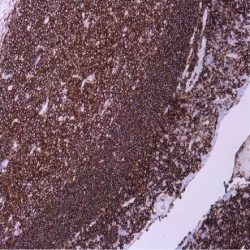
آنتی بادی CD43 (DF-T1)
Name: CD43 Antibody Clone DF-T1
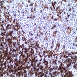
Description and aplications: CD43 (leukosialin, sialophorin, or leukocyte sialoglycoprotein) is a cell surface glycoprotein which is expressed on all thymocytes and T-cells. CD43 is involved in activation of T cells, B cells, NK cells, and monocytes. The anti-CD43 antibody recognizes a cell surface glycoprotein of 95/115/135kDa (depending upon the extent of glycosylation), identified as CD43. 70-90% of T-cell lymphomas and from 22-37% of B-cell lymphomas express CD43. No reactivity has been observed with reactive B-cells. So a B-lineage population that co-expresses CD43 is highly likely to be a malignant lymphoma, especially a low-grade lymphoma, rather than a reactive B-cell population. When CD43 antibody is used in combination with anti-CD20, effective immunophenotyping of the lymphomas in formalin-fixed tissues can be obtained. Co-staining of a lymphoid infiltrate with anti-CD20 and anti-CD43 argues against a reactive process and favors a diagnosis of lymphoma.
Composition: anti-human CD43 mouse monoclonal antibody purified from ascites. Prepared in 10mM PBS, pH 7.4, with 0.2% BSA and 0.09% sodium azide.
Immunogen: Myeloblastic KG1 cells.
-
 آنتی بادیهای ایمونوهیستوشیمی
آنتی بادی CD45 (2B11&PD7/26)
Name: CD45 (leukocyte common antigen – LCA)Antibody (cocktail of clones 2B11 & PD7/26)
Description and aplications: Anti-CD45 (anti-leukocyte common antigen) is routinely used to aid the differential diagnosis of undifferentiated neoplasms, whenever malignant lymphoma is suspected by the morphological or clinical data. It is a highly specific antibody, therefore a positive result is highly indicative of hematolymphoid origin. Certain types of hematolymphoid neoplasms may lack CD45 (Hodgkin lymphoma, some T-cell lymphomas, and some leukemias) so its absence does not rule out a hematolymphoid tumor. This antibody is expressed almost exclusively by cells of hematopoietic lineage and is present in most benign and malignant lymphocytes as well as plasma cell precursors.
Composition: anti-human CD45 mouse monoclonal antibody purified from ascites fluid by Protein A chromatography. Prepared in 10mM PBS, pH 7.4, with 0.2% BSA and 0.09% sodium azide.
-
 آنتی بادیهای ایمونوهیستوشیمی
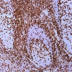
آنتی بادی CD45RO (UCHL-1)
Name: CD45RO Antibody Clone UCHL-1
Description and applications: The CD45RO antibody recognizes a 180-185kDa protein, identified as an isoform of leukocyte common antigen (CD45RO) (4th Leucocyte Typing Workshop: Code No. N31).The epitope recognized by this antibody is sensitive to neuraminidase digestion. This antibody reacts with mature activated T-cells, most thymocytes, and a sub-population of resting T-cells
within both CD4 and CD8 subsets. It shows no reactivity with normal B or natural killer cells, but reacts with granulocytes and monocytes. Reportedly, it is useful to identify T-cell lymphomas and leukemias. It rarely stains NK cells or B-cell lymphomas.Composition: Anti-human CD45RO mouse monoclonal antibody purified from serum and prepared in 10mM PBS, pH 7.4, with 0.2% BSA and 0.09% sodium azide.
Immunogen: Cultured human T-cells from an IL-2- dependent T-cell line (CA1).
-
 آنتی بادیهای ایمونوهیستوشیمی
آنتی بادی CD45RB (BRA-11)
Name: CD45RB Antibody Clone BRA-11
Description and applications: CD45 has been identified as a transmembrane glycoprotein, broadly expressed among hematopoietic cells. Multiple isoforms of CD45 are distributed through-out the immune system according to cell type. These isoforms arise because of alternative splicing of exons 4, 5 and 6. The corresponding protein domains are characterized by the binding of monoclonal antibodies specific for CD45RA (exon 4), CD45RB (exon 5), CD45RC (exon 6) and CD45RO (exons 4 to 6 spliced out). The variation in these isoforms is localized to the extracellular domain of CD45, while the intracellular domain is conserved. CD45 functions as a phosphotyrosine phosphatase, a vital component for efficient tyrosine phosphorylation induction by the TCR/CD3 complex. The tyrosine phosphatase activity of CD45 is contained within the conserved intracellular domain. Src and Syk family protein tyrosine kinases are utilized by the TCR/CD3 complex to initiate signaling cascades. Several members of these two families, including Lck, Fyn and ZAP-70, have been implicated as physiological substrates of CD45. CD45RB is expressed in a high proportion of circulating B lymphocytes, suppressor and cytotoxic T lymphocytes, a subpopulation of helper T lymphocytes and in most thymocytes. Expression of CD45RB decreases as the T cells mature from naïve to memory mature T cells, suggesting that its expression is predominantly in immature T lymphocytes. The anti-CD45RB antibody is useful in the study of lymphocyte subpopulations in autoimmune diseases, especially inflammatory bowel disease, and for the immunophenotypic characterization of leukemias and lymphomas.
Composition: Anti-human CD45RB mouse monoclonal antibody purified from serum and prepared in 10mM PBS, pH 7.4, with 0.2% BSA and 0.09% sodium azide.
-
 آنتی بادیهای ایمونوهیستوشیمی
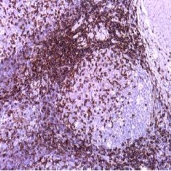
آنتی بادی CD5 (EP77)
Name: CD5 Antibody Clone EP77
Description and applications:CD5 (Lymphocyte antigen T1/Leu-1) is a transmembrane glycoprotein which has been implicated as a receptor in the regulation of T-cell proliferation. CD5 antibody labels a variety of T lymphocytes, mantle zone lymphocytes and a small subset of B lymphocytes. In tumors, CD5 is expressed on T-cell malignancies, B cell chronic lymphocytic leukemia (CLL)/small lymphocytic lymphoma (SLL), and mantle-cell lymphoma. As CD5 is expressed in activated T lymphocytes and in several T-cell leukemias, it must be included in the panel of immunophenotypic study of T cell lymphomas as it is positive in mycosis fungoides and is deleted in most Tcell anaplastic lymphomas (ALK), lymphomatoid papulosis, and blastoid NK / T lymphomas. In addition, anti-CD5 is helpful in diagnosis of thymic carcinoma (CD5 positive).
Composition: Anti-human CD5 rabbit monoclonal antibody purified from serum and prepared in 10mM PBS, pH 7.4, with 0.2% BSA and 0.09% sodium azide.
IMMUNOGEN: A synthetic peptide corresponding to
residues in human CD5 protein. -
 آنتی بادیهای ایمونوهیستوشیمی
آنتی بادیCD54/ICAM -کلون23G12
Name: CD54/ICAM-1 Monoclonal Antibody Clone 23G12
Description and applications: ICAM-1 (intercellular adhesion molecule) has 7 potential N-linked glycosylation sites. It is a single
chain glycoprotein of Ig supergene family, present on unstimulated endothelial cells (EC) and on a variety of other cell types including activated fibroblasts, EC, macrophages, and lymphocytes. ICAM-1 mediates cell adhesion by binding to integrins CD11a/CD18 (leukocyte adhesion molecule, LFA-1) and to CD11b/CD18 (Mac-1).Composition: Anti-human CD54/ICAM-1 mouse monoclonal antibody purified from serum and prepared in 10mM PBS, pH 7.4, with 0.2% BSA and 0.09% sodium azide.
Immunogen:Recombinant protein encoding part of the external domain of human CD54.
-
 آنتی بادیهای ایمونوهیستوشیمی
آنتی بادی CD56/NCAM-1 (123C3)
Name: CD56/NCAM-1 Antibody Clone 123C3
Description and applications: CD56, known as neural cell adhesion molecule (NCAM), was originally identified in the nervous system and belongs to a group of cell adhesion molecules including cadherins, selectins, and integrins. Anti-CD56 recognizes two proteins of the NCAM, the basic molecule expressed on most neuroectodermally-derived cell lines, tissues and neoplasms (e.g. neuroblastomas, small cell carcinomas). It is also expressed on some mesodermally-derived tumors (rhabdomyosarcoma). Furthermore, anti-CD56 has found great utility in the recognition of NK cell and NK/T-cell lymphomas. It has been shown that 71% of myelomas are positive for anti-CD56 as well. It also offers higher sensitivity in the diagnosis of small cell carcinoma than anti-chromogranin and anti-synaptophysin and has been demonstrated as a useful marker for sex cord gonadal stromal tumors. Light staining of smooth muscle elements may be seen.
Composition: anti-human CD56 rabbit monoclonal antibody purified from ascites. Prepared in 10mM PBS, pH 7.4, with 0.2% BSA and 0.09% sodium azide.
-
 آنتی بادیهای ایمونوهیستوشیمی
آنتی بادی CD57 (NK-1)
Name: CD57 Antibody Clone NK-1
Description and applications: CD57 is expressed on a subpopulation of 15-20% of peripheral blood mononuclear cells, about 60% of NK active cells and on a subset of T cells. It also reacts with a variety of cell types in non-lymphoid tissues. Staining was observed in glandular epithelium of prostate, peripheral nerve and central nervous system, glandular epithelium of ovary, bronchiolar epithelium and in syncytiotrophoblasts. Variable staining was seen in the colloid of thyroid and some focal staining in kidney epithelium. Additionally, some staining was observed in capillaries of various tissues, subsets of pancreatic islet cells, hepatocytes and ovarian stromal cells. It stains neuroendocrine cells and their tumors. Identification of natural killer cells (NK) in tissue sections or cell suspensions together with other lymphoid markers may be useful in identifying indolent lymphoproliferative disorders of NK cells (CD57 +), leukemias large granular T cells (CD57 is expressed in the common variant), NK / T extranodal lymphoma (usually CD56 + / CD57-) and Hodgkin lymphoma nodular lymphocyte-predominant (nodular paragranuloma) which contains an abundant reactive population of cells that frequently surrounds the tumour cells (rosette-like). Additional staining was observed in a range of common tumours including colon adenocarcinoma, prostatic adenocarcinoma and ovarian carcinoma.
Composition: anti-human CD57 mouse monoclonal antibody purified from ascites and prepared in 10mM PBS, pH 7.4, with 0.2% BSA and 0.09% sodium azide.
Immunogen: Human peripheral blood mononuclear cells.
-
 آنتی بادیهای ایمونوهیستوشیمی
آنتی بادی CD63 (NKI/C3)
Name: CD63 Antibody Clone NKI/C3
Description and applications:The CD63 molecule, also called melanoma-associated antigen MLA1 or ME491, granulophysine, or
membrane-associated lysosomal glycoprotein 3 (LAMP-3), is a 53-kDa glycoprotein belonging to the transmembrane 4 protein superfamily or tetraspanins. It is made up of 237 amino acids and is commonly expressed in melanoma tumour cells, where it is associated with early stages of tumour progression. The ME491 gene, which encodes it, is located on the chromosomal region 12q13.2 and the lack of this protein has been observed in Hermansky- Pudlak syndrome, which is characterized by the presence of deficient lysosomes with dense granules and accumulation of ceroid material in reticuloendothelial system cells. Functionally, CD63 plays an important role in intracellular transport and is a necessary molecule in the trafficking processes that take place in the PMEL luminal domain, which are essential in the development and maturation of melanocytes. Similarly, CD63 is important for leukocyte-endothelial cell adhesion through activation by P-selectin. Finally, CD63 appears to be involved in degranulation of mast cells in response to their activation through subunit beta of high affinity immunoglobulin receptor’s epsilon chain (Ms4a2/FceRI system). CD63 antigen is widely distributed in normal tissues,being present in platelets’ lysosomal granules, neutrophil granulocytes, and basophil granulocytes, as well as in a small proportion of inactive T cells, endothelial cells, and macrophages. In tumour cells, CD63 is expressed in dysplastic nevi and radial growth phase melanomas, as well as in renal angiomyolipoma, cellular neurothekeoma, atypical fibroxanthoma and dermatofibrosarcoma protuberans, clear cell fibrous papule of the nose, and non-neural granular cell tumour of the skin. Its usefulness has also been proven in the differential diagnosis between renal oncocytomas, which show apical staining, and renal cell carcinomas with eosinophilic cytoplasm, which show diffuse cytoplasmic staining. Other tumours such as breast carcinomas, Merkel cell carcinomas, astrocytomas, and lung adenocarcinomas may also show positive staining, being, in the latter two cases, an indicator of a better prognosis.Composition: Anti-human CD63 mouse monoclonal antibody purified from serum and prepared in 10mM PBS, pH 7.4, with 0.2% BSA and 0.09% sodium azide.
-
 آنتی بادیهای ایمونوهیستوشیمی
آنتی بادی CD7 (EP132)
Name: CD7 Antibody-Clone-EP132
Description and aplications: This antibody recognizes a glycoprotein of 40kD molecular weight known as CD7 (also known as TP40 and Leu9). CD7 is positioned at 17q25.2-q25.3 and present on thymocytes, mature T cells, and NK cells. During T cell differentiation, CD7 is one of the earliest lineage markers to appear, therefore, the marker is considered the most clinically useful for acute lymphoid leukemia of T origin. In contrast, the CD7 molecule is absent in some cases as severe combined immunodeficiency. As the CD7 antigen is expressed in mature and immature T cells and NK cells, including those with mixed immuophenotype myeloid (precursor NK/myeloid cells leukemia), the anti-CD7 antibody is useful for the identification of lymphoid neoplasms derived therefrom. However it should be noted that in the peripheral T-cell lymphomas, frequent deletion or pre- and post-transcriptional regulation can lead to the loss of CD7 expression in the cell membrane, therefore their absence in this case does not rule out the origin of the T neoplasia. It is also not unusual in benign inflammatory dermatoses that reactive T cells do not express CD7, therefore, these histopathological features must be considered in the differential diagnosis.
Composition: anti-CD7 rabbit monoclonal antibody obtained from supernatant culture and prediluted in a tris buffered solution pH 7.4 containing 0.375mM sodium azide solution as bacteriostatic and bactericidal.
Immunogen: A synthetic peptide corresponding to residues of human CD7 protein.
-
 آنتی بادیهای ایمونوهیستوشیمی
آنتی بادی CD71 (10F11)
Name: CD71 Antibody Clone 10F11
Description and aplications: CD71 recognizes a 90kDa glycoprotein encoded by a gene located in the chromosomal region3q29. It is a recirculating molecule between extra- and intracellular environment, located on the surface of all proliferating cells, which has an important role in the transcellular transport of iron. It is specifically expressed on the surface of the precursor cells of erythroid line, with reduced expression in reticulocytes and complete loss of expression in mature erythroid elements. In normal tissues is a useful marker in identifying erythroid precursors or dispoyétic cells of the normal bone marrow as the other cells of myeloid lineage or mature erythroid cells are negative. Other cells that can normally express the antibody are placental syncytiotrophoblasts, myocytes, basal keratinocytes, hepatocytes, cells of pancreatic Langerhans islet and spermatocytes. In tumoral lesions CD71 is a marker with diagnostic value for cases of erythroid leukemia, while the dysplastic erythroid precursors have a decreased expression. In all these cases, compared with the staining of Anti-Haemoglobin and glycophorin A antibodies, CD71 is more sensitive and specific for not presenting reaction with the mature erythroid elements, facilitating the interpretation. Similar results are obtained in biopsies fixed in Zenker’s fixative or paraffin. Except erythroid leukemias, the antibody is negative in all primary tumors or metastatic bone marrow lessions.
In extramedullary lymphomas, the antibody is positive in isolated cases of diffuse large B-cell lymphoma, peripheral T lymphomas, large cell anaplastic lymphomas and in the Reed-Sternberg cells of Hodgkin lymphoma. In breast carcinomas, CD71 expression was detected in a group of tumours with luminal and basal genotype (more common in medullary carcinoma) resistant to treatment with Tamoxifen. In experimental studies on breast carcinoma, models using antibodies directed against CD71, an inhibition of cell proliferation and survival has been observed, suggesting CD71 as a possible new therapeutic target in cases of CD71 positive carcinoma.Composition: anti-human CD71 mouse monoclonal antibody purified from ascites. Prepared in 10mM PBS, pH 7.4, with 0.2% BSA and 0.09% sodium azide.
Immunogen: Prokaryotic recombinant protein corresponding to a region of the N terminal intracellular domain of Transferrin Receptor (Human).
برای تماس کلیک کنید
سما تشخیص آریا ، عرضه کننده متنوع ترین و کامل ترین محصولات کیت های آزمایشگاهی