-
 آنتی بادیهای ایمونوهیستوشیمی
آنتی بادی CD57 (NK-1)
Name: CD57 Antibody Clone NK-1
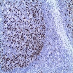
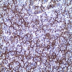
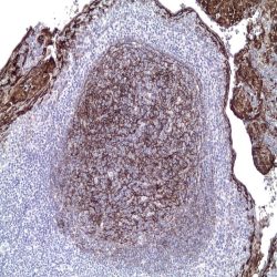
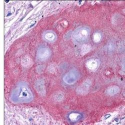
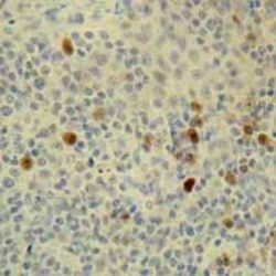
Description and applications: CD57 is expressed on a subpopulation of 15-20% of peripheral blood mononuclear cells, about 60% of NK active cells and on a subset of T cells. It also reacts with a variety of cell types in non-lymphoid tissues. Staining was observed in glandular epithelium of prostate, peripheral nerve and central nervous system, glandular epithelium of ovary, bronchiolar epithelium and in syncytiotrophoblasts. Variable staining was seen in the colloid of thyroid and some focal staining in kidney epithelium. Additionally, some staining was observed in capillaries of various tissues, subsets of pancreatic islet cells, hepatocytes and ovarian stromal cells. It stains neuroendocrine cells and their tumors. Identification of natural killer cells (NK) in tissue sections or cell suspensions together with other lymphoid markers may be useful in identifying indolent lymphoproliferative disorders of NK cells (CD57 +), leukemias large granular T cells (CD57 is expressed in the common variant), NK / T extranodal lymphoma (usually CD56 + / CD57-) and Hodgkin lymphoma nodular lymphocyte-predominant (nodular paragranuloma) which contains an abundant reactive population of cells that frequently surrounds the tumour cells (rosette-like). Additional staining was observed in a range of common tumours including colon adenocarcinoma, prostatic adenocarcinoma and ovarian carcinoma.
Composition: anti-human CD57 mouse monoclonal antibody purified from ascites and prepared in 10mM PBS, pH 7.4, with 0.2% BSA and 0.09% sodium azide.
Immunogen: Human peripheral blood mononuclear cells.
-
 آنتی بادیهای ایمونوهیستوشیمی
آنتی بادی CD63 (NKI/C3)
Name: CD63 Antibody Clone NKI/C3
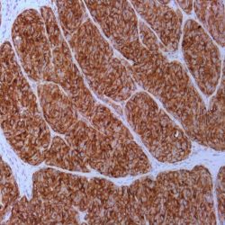
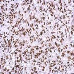
Description and applications:The CD63 molecule, also called melanoma-associated antigen MLA1 or ME491, granulophysine, or
membrane-associated lysosomal glycoprotein 3 (LAMP-3), is a 53-kDa glycoprotein belonging to the transmembrane 4 protein superfamily or tetraspanins. It is made up of 237 amino acids and is commonly expressed in melanoma tumour cells, where it is associated with early stages of tumour progression. The ME491 gene, which encodes it, is located on the chromosomal region 12q13.2 and the lack of this protein has been observed in Hermansky- Pudlak syndrome, which is characterized by the presence of deficient lysosomes with dense granules and accumulation of ceroid material in reticuloendothelial system cells. Functionally, CD63 plays an important role in intracellular transport and is a necessary molecule in the trafficking processes that take place in the PMEL luminal domain, which are essential in the development and maturation of melanocytes. Similarly, CD63 is important for leukocyte-endothelial cell adhesion through activation by P-selectin. Finally, CD63 appears to be involved in degranulation of mast cells in response to their activation through subunit beta of high affinity immunoglobulin receptor’s epsilon chain (Ms4a2/FceRI system). CD63 antigen is widely distributed in normal tissues,being present in platelets’ lysosomal granules, neutrophil granulocytes, and basophil granulocytes, as well as in a small proportion of inactive T cells, endothelial cells, and macrophages. In tumour cells, CD63 is expressed in dysplastic nevi and radial growth phase melanomas, as well as in renal angiomyolipoma, cellular neurothekeoma, atypical fibroxanthoma and dermatofibrosarcoma protuberans, clear cell fibrous papule of the nose, and non-neural granular cell tumour of the skin. Its usefulness has also been proven in the differential diagnosis between renal oncocytomas, which show apical staining, and renal cell carcinomas with eosinophilic cytoplasm, which show diffuse cytoplasmic staining. Other tumours such as breast carcinomas, Merkel cell carcinomas, astrocytomas, and lung adenocarcinomas may also show positive staining, being, in the latter two cases, an indicator of a better prognosis.Composition: Anti-human CD63 mouse monoclonal antibody purified from serum and prepared in 10mM PBS, pH 7.4, with 0.2% BSA and 0.09% sodium azide.
-
 آنتی بادیهای ایمونوهیستوشیمی
آنتی بادی CD7 (EP132)
Name: CD7 Antibody-Clone-EP132
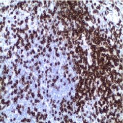
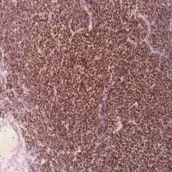
Description and aplications: This antibody recognizes a glycoprotein of 40kD molecular weight known as CD7 (also known as TP40 and Leu9). CD7 is positioned at 17q25.2-q25.3 and present on thymocytes, mature T cells, and NK cells. During T cell differentiation, CD7 is one of the earliest lineage markers to appear, therefore, the marker is considered the most clinically useful for acute lymphoid leukemia of T origin. In contrast, the CD7 molecule is absent in some cases as severe combined immunodeficiency. As the CD7 antigen is expressed in mature and immature T cells and NK cells, including those with mixed immuophenotype myeloid (precursor NK/myeloid cells leukemia), the anti-CD7 antibody is useful for the identification of lymphoid neoplasms derived therefrom. However it should be noted that in the peripheral T-cell lymphomas, frequent deletion or pre- and post-transcriptional regulation can lead to the loss of CD7 expression in the cell membrane, therefore their absence in this case does not rule out the origin of the T neoplasia. It is also not unusual in benign inflammatory dermatoses that reactive T cells do not express CD7, therefore, these histopathological features must be considered in the differential diagnosis.
Composition: anti-CD7 rabbit monoclonal antibody obtained from supernatant culture and prediluted in a tris buffered solution pH 7.4 containing 0.375mM sodium azide solution as bacteriostatic and bactericidal.
Immunogen: A synthetic peptide corresponding to residues of human CD7 protein.
-
 آنتی بادیهای ایمونوهیستوشیمی
آنتی بادی CD71 (10F11)
Name: CD71 Antibody Clone 10F11
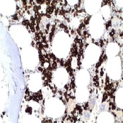
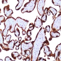
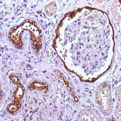
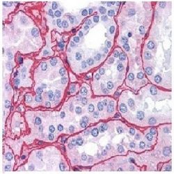
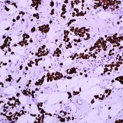
Description and aplications: CD71 recognizes a 90kDa glycoprotein encoded by a gene located in the chromosomal region3q29. It is a recirculating molecule between extra- and intracellular environment, located on the surface of all proliferating cells, which has an important role in the transcellular transport of iron. It is specifically expressed on the surface of the precursor cells of erythroid line, with reduced expression in reticulocytes and complete loss of expression in mature erythroid elements. In normal tissues is a useful marker in identifying erythroid precursors or dispoyétic cells of the normal bone marrow as the other cells of myeloid lineage or mature erythroid cells are negative. Other cells that can normally express the antibody are placental syncytiotrophoblasts, myocytes, basal keratinocytes, hepatocytes, cells of pancreatic Langerhans islet and spermatocytes. In tumoral lesions CD71 is a marker with diagnostic value for cases of erythroid leukemia, while the dysplastic erythroid precursors have a decreased expression. In all these cases, compared with the staining of Anti-Haemoglobin and glycophorin A antibodies, CD71 is more sensitive and specific for not presenting reaction with the mature erythroid elements, facilitating the interpretation. Similar results are obtained in biopsies fixed in Zenker’s fixative or paraffin. Except erythroid leukemias, the antibody is negative in all primary tumors or metastatic bone marrow lessions.
In extramedullary lymphomas, the antibody is positive in isolated cases of diffuse large B-cell lymphoma, peripheral T lymphomas, large cell anaplastic lymphomas and in the Reed-Sternberg cells of Hodgkin lymphoma. In breast carcinomas, CD71 expression was detected in a group of tumours with luminal and basal genotype (more common in medullary carcinoma) resistant to treatment with Tamoxifen. In experimental studies on breast carcinoma, models using antibodies directed against CD71, an inhibition of cell proliferation and survival has been observed, suggesting CD71 as a possible new therapeutic target in cases of CD71 positive carcinoma.Composition: anti-human CD71 mouse monoclonal antibody purified from ascites. Prepared in 10mM PBS, pH 7.4, with 0.2% BSA and 0.09% sodium azide.
Immunogen: Prokaryotic recombinant protein corresponding to a region of the N terminal intracellular domain of Transferrin Receptor (Human).
-
 آنتی بادیهای ایمونوهیستوشیمی
آنتی بادی CD99 (EP8)
Name: CD99 Antibody Clone EP8
Description and applications: CD99 is a transmembrane glycoprotein, also known as MIC2. It is involved in T cell adhesion, leukocyte migration and differentiation of primitive neuroectodermal cell.
CD99 labels lymphocyte, ovarian granulosa cells, pancreatic islet cells, Sertoli cells, CNS ependymal cells and endothelial cells. CD99 has been useful in diagnosis of Ewing’s sarcoma, sex cord-stromal tumor, endocrine tumor of pancreas. Additionally, CD99 is found in a subset of other tumors including lymphoblastic lymphoma, breast carcinoma and other malignancies.
This CD99 antibody has been validated with excellent staining result by Nordic Immunohistochemical Quality Control (NordiQC).Composition: anti-human CD99 rabbit monoclonal antibody purified by protein affinity. Prepared in 10mM PBS, pH 7.4, with 0.2% BSA and 0.09% sodium azide.
Immunogen: A synthetic peptide corresponding to residues on the C-terminus of human CD99 protein.
-
 آنتی بادیهای ایمونوهیستوشیمی
آنتی بادی CDX-2 (EP25)
Name: CDX2 Antibody Clone EP25
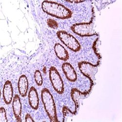
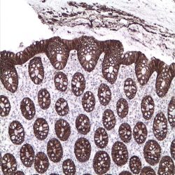
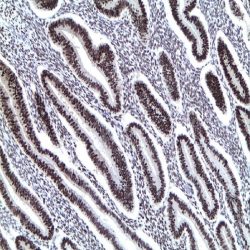
Description and applications: The caudal-related homeodomain protein 2, CDX-2, is a transcription factor which is expressed in the intestine and is thought to play an important role in the proliferation and differentiation of intestinal epithelial cells. The CDX-2 protein is expressed in primary and metastatic colorectal carcinomas, intestinal metaplasia of the stomach and intestinal type gastric cancer. In human colorectal cancer, the expression of both CDX2 and carbonic anhydrase 1, a gene regulated by CDX2, is reduced or absent. CDX-2 is one of the important regulators in defining pathways for coordinate control of drug metabolism in the gastrointestinal tract. Most of the mucinous adenocarcinomas of the ovary, ampullary, gastric or esophageal adenocarcinomas show immunoreactivity for CDX2 even though,heterogeneous and less intense than in colorectal carcinomas. In endometrial and pancreatic adenocarcinomas CDX2 expression it is exceptional while breast adenocarcinomas, including the mucinous ones, stain negative for CDX2. Approximately 10% of lung adenocarcinomas may be positive for CDX2.
Composition: anti-human Thyroglobulin rabbit monoclonal antibody purified from ascites. Prepared in 10mM PBS, pH 7.4, with 0.2% BSA and 0.09% sodium azide.
Immunogen: A synthetic peptide corresponding to residues near the C-term of human CDX-2 protein.
Name: CDX2 Antibody (Clone EP25)
Description and applications: The caudal-related homeodomain protein 2, CDX-2, is a transcription factor which is expressed in the intestine and is thought to play an important role in the proliferation and differentiation of intestinal epithelial cells. The CDX-2 protein is expressed in primary and metastatic colorectal carcinomas, intestinal metaplasia of the stomach and intestinal type gastric cancer. In human colorectal cancer, the expression of both CDX2 and carbonic anhydrase 1, a gene regulated by CDX2, is reduced or absent. CDX-2 is one of the important regulators in defining pathways for coordinate control of drug metabolism in the gastrointestinal tract. Most of the mucinous adenocarcinomas of the ovary, ampullary, gastric or esophageal adenocarcinomas show immunoreactivity for CDX2 even though,heterogeneous and less intense than in colorectal carcinomas. In endometrial and pancreatic adenocarcinomas CDX2 expression it is exceptional while breast adenocarcinomas, including the mucinous ones, stain negative for CDX2. Approximately 10% of lung adenocarcinomas may be positive for CDX2.
Composition: anti-human Thyroglobulin rabbit monoclonal antibody purified from ascites. Prepared in 10mM PBS, pH 7.4, with 0.2% BSA and 0.09% sodium azide.
Immunogen: A synthetic peptide corresponding to residues near the C-term of human CDX-2 protein.
-
 آنتی بادیهای ایمونوهیستوشیمی
آنتی بادی CGA (Glycoprotein Hormone Alpha) (EP3373)
Name : Rabbit anti-human CGA (Glycoprotein Hormone Alpha) Monoclonal Antibody clone EP3373
Description and applications: CGA belongs to the glycoprotein hormones alpha chain family, which includes the alpha subunits of chorionic gonadotropin (CG), luteinizing hormone (LH), follicle-stimulating hormone (FSH), and thyroidstimulating hormone (TSH). Glycoprotein hormones have the most complex chemical structures of the classical protein hormones. They are dimers consisting of alpha and beta subunits that are covalently associated. These hormones share the same alpha subunit; however, their beta chains are unique and confer them biological specificity. These hormones are essential for the complex endocrine system, which regulates normal growth, sexual development, and reproductive function.The gene encoding the chorionic gonadotropin alpha chain is located on the chromosome region 6q14.3. Hormones LH, FSH, and TSH are secreted by the anterior pituitary, whereas CG is secreted by the placenta. Immunohistochemical detection of chorionic gonadotropin alpha and beta subunits is observed in most non-functional pituitary adenomas, whereas only about 0.6% of these adenomas express the alpha subunit exclusively. In serum, hypersecretion of chorionic gonadotropin alpha subunit is observed in more than 65% of non-functional adenomas, which makes this antibody a useful tool for confirming diagnosis.
Composition: Anti-human CGA rabbit monoclonal antibody purified from serum and prepared in 10mM PBS, pH 7.4, with 0.2% BSA and 0.09% sodium azide
Intended use: Immunohistochemistry (IHC) on paraffin embedded tissues. Not tested on frozen tissues or Western-Blotting
Immunogen: Synthetic peptide corresponding to human protein CGA residues.
-
 آنتی بادیهای ایمونوهیستوشیمی
آنتی بادی Chromogranin A (LK2H10)
Name: Chromogranin A Antibody Clone LK2H10
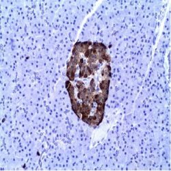
Description and aplications: Chromogranin A (a protein of 439-amino acid which is encoded on chromosome 14) is present in neuroendocrine cells throughout the body, including the neuroendocrine cells of the large and small intestine, adrenal medulla and pancreatic islets. It is an excellent marker for carcinoid tumors, pheo-chromocytomas, paragangliomas, and other neuro-endocrine tumors. Coexpression of chromogranin A and neuron specific enolase (NSE) is common in neuroendocrine neoplasms.
Composition: anti-human Chromogranin A mouse monoclonal antibody purified from ascites. Prepared in10mM PBS, pH 7.4, with 0.2% BSA and 0.09% sodium azide.
Immunogen: Human pheochromocytoma.
-
 آنتی بادیهای ایمونوهیستوشیمی
آنتی بادی Clusterin (EP181)
Name: Clusterin Antibody Clone EP181
Description and applications: This monoclonal antibody recognises clusterin, a heterodimeric glycoprotein of 70-80 kDa. Clusterin is highly expressed in various tissues including pancreas, tonsil, nasopharynx, uterine cervix, and epididymis; it is also secreted into different fluids. Human clusterin is encoded by a single copy gene located on chromosome 8p12 and 8p21 with nine exons and eight introns, spanning approximately 17 kb. Clusterin has two isoforms: one is secreted directly into different fluids, and the other is expressed in the cytoplasm and in the cell nucleus. Clusterin is involved in numerous biological processes including lipid transport, epithelial differentiation, cell-matrix interaction, cell aggregation, and regulation of apoptosis in hormone-dependent epithelia. Clusterin has recently been proposed as an immunohistochemical marker specific for follicular dendritic cell tumours, since it is not expressed in the rest of dendritic cell neoplasms (Langerhans cell histiocytosis and interdigitating dendritic cell tumours) and it is very useful when included in the panel comprising CD21, CD35, S-100 protein, CD1a, CD68, actin, desmin, and cytokeratin for the differential diagnosis of dendritic cell tumours. Clusterin gene expression has been demonstrated by microarray analysis in systemic (80-100% of cases) and cutaneous (40-60%) anaplastic lymphomas, in some large B-cell lymphomas (10%), and very sporadically in peripheral T lymphomas and nodular sclerosis Hodgkin lymphomas. Among non-hematopoietic neoplasms, clusterin overexpression has been detected in breast, colon, pancreas, and prostate carcinomas. Overexpression in prostate carcinomas inhibits neoplastic cells’ apoptotic death; therefore, a protective effect against cell death by apoptosis is associated to this protein.
Composition: Anti-human Clusterin rabbit monoclonal antibody purified from serum and prepared in 10mM PBS, pH 7.4, with 0.2% BSA and 0.09% sodium azide.
Immunogen: Synthetic peptide corresponding to residues from human clusterin.
-
 آنتی بادیهای ایمونوهیستوشیمی
آنتی بادی Claudin-1 (Polyclonal)
Name: Claudin-1 Antibody (Polyclonal)
Description and applications: Claudin proteins are a family of proteins associated with tight junctions. Tight junctions are specialized regions of cell to cell contact; made up of network of strands to act as a molecular gasket for preventing the leakage of ions, water etc. between cells. They are abundant in luminal epithelial sheets where they maintain epithelial cell polarity. Different tissues exhibit different Claudin composition.
Composition: anti-human Claudin-1 rabbit polyclonal antibody purified from serum and prepared in 10mM PBS, pH 7.4, with 0.2% BSA and 0.09% sodium azide
Immunogen: Synthetic peptide corresponding to the C-terminus of human Claudin 1.
-
 آنتی بادیهای ایمونوهیستوشیمی
آنتی بادی Claudin-7 (Polyclonal)
Name: Claudin-7 Antibody (Polyclonal)
Description and applications: The claudin protein family consists of 24 different cellcell adhesion molecules that constitute the fundamental core of tight junctions, which are in charge of controlling the molecule flow between epithelial cells. This control is performed by creating a biochemical barrier against water and restricting lateral diffusion of membrane lipids and proteins towards the paracellular spaces between the apical and the basement membrane, which, however, allows polar molecule transport. This antibody recognises claudin 7, whose expression is controlled by a gene located on chromosome 17. In addition to the general metabolic control functions described above for claudins, claudin 7, specifically in its phosphorylated form: 1) interacts with EPCAM by regulating its expression; 2) promotes E-cadherin normal expression by epithelial cells; and 3) regulates the expression of prostate-specific antigen (PSA) and is involved in the androgen receptor regulation mechanism. In this sense, its maximum expression takes place in tissues with normal levels of dihydrotestosterone (DHT), while high concentrations of this molecule inhibit its presence. Claudin 7 is frequently expressed in the distal nephron epithelium, prostate, airway epithelium, and other glandular-type epithelia where it shows membrane staining. CNS cells and lymphoid cells are negative. Claudins are present in a variety of normal, hyperplastic, and benign tissues as well as in benign and malignant neoplasms that show epithelial differentiation. It is for this reason that the differential expression of its different members can be used to confirm the histological identity of some neoplasms. Thus, claudin immunohistochemical detection, including claudin 7, makes it possible to differentiate and confirm the diagnosis between: 1) oncocytomas and chromophobe cell renal carcinomas; 2) endometrioid carcinoma and uterine papillary serous carcinoma; 3) mesothelioma and adenocarcinoma metastasis; 4) hepatocellular carcinoma and cholangiocarcinoma; and 5) diffuse and intestinal-type gastric carcinomas. Its usefulness has also been shown for distinguishing between highgrade ductal in situ and lobular carcinomas, and infiltrating carcinomas of the breast. In addition, its cytoplasmic expression in solid pseudopapillary tumours of the pancreas enables the differential diagnosis with other tumours such as pancreatoblastoma, acinar cell carcinoma, or pancreatic neuroendocrine tumours since all of them show membrane staining for claudin 7. Similarly, the expression of certain claudins can be used as a prognostic marker in various neoplasms such as nasopharyngeal carcinomas, where claudin 7 overexpression correlates with tumour grade, presence of distant metastases, and survival. Similar results have been obtained in ovarian adenocarcinomas. Likewise, an increase in the expression of claudins 1 and 7 has also been detected in cervical premalignant lesions and invasive neoplasms compared to normal epithelium.
Composition: anti-human Claudin-7 rabbit polyclonal antibody purified from serum and prepared in 10mM PBS, pH 7.4, with 0.2% BSA and 0.09% sodium azide.
Immunogen: Synthetic peptide corresponding to the C-terminal domain of human claudin 7.
-
 آنتی بادیهای ایمونوهیستوشیمی
آنتی بادی C-Myc (Y69)
Name: c-Myc Antibody clone Y69
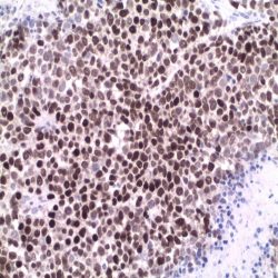
Description and applications: The c-MYC gene is located at chromosome 8q24. It is required for
progression through the cell cycle and promotes cellular proliferation. The t(8;14)(q24;q32) translocation and the c-MYC/immunoglobulin heavy-chain (IGH) fusion gene are not only in Burkitt lymphoma, but are also seen in diffuse large B-cell lymphoma, blastic mantle cell lymphoma and transformed follicular lymphoma. In another study on predicting c-MYC translocation in 17 cases of Burkitt lymphomas (BLs) and 19 cases of diffuse large B-cell lymphomas (DLBCLs), Ruzinova et al. reported that the sensitivity and specificity of this c-Myc antibody on identifying tumor harboring a c- MYC rearrangement reached 96% and 90% respectively. This novel c-Myc antibody is a useful tool for identifying aggressive B-cell lymphomas likely to harbor a c-MYC rearrangement, and thus warrant genetic testing. In addition to its aberrant expression in lymphomas, c-Myc amplification and overexpression are also implicated in tumor progression or prognosis in many other malignancies including pancreatic cancer, breast cancer, prostate cancer, bladder cancer and soft tissue leiomyosarcoma. By utilizing this anti-c- Myc antibody coupled with genetically defined control experiments, a recent study by Gurel et al. demonstrated the specificity of this c-Myc antibody in the staining of paraffin-embedded prostate normal and tumor tissues. Upregulation of nuclear c-Myc protein expression may be a critical oncogenic event in driving human prostate cancer initiation and progression.Composition: Anti-human c-Myc rabbit monoclonal antibody purified from serum and prepared in 10mM PBS, pH 7.4, with 0.2% BSA and 0.09% sodium azide.
Immunogen: A synthetic peptide corresponding to residues in N-terminus of human c-Myc protein.
-
 آنتی بادیهای ایمونوهیستوشیمی
آنتی بادی Collagen II (2B1.5)
Name: Collagen II Antibody Clone 2B1.5
Description and applications:This antibody is highly specific to type II collagen and shows no crossreaction with types I, III, IV, V, VI, IX, X, or XI. In Western blotting, 2B1.5 MAb reacts with the TCB fragment of pepsin digested type II collagen after digestion with mammalian collagenase. It also reacts with lathyritic type II collagen.
Composition: Anti-human Collagen II mouse monoclonal antibody purified from serum and prepared in 10mM PBS, pH 7.4, with 0.2% BSA and 0.09% sodium azide.
Immunogen: A purified preparation of lathyritic type II collagen from embryonic chicken sternum.
-
 آنتی بادیهای ایمونوهیستوشیمی
آنتی بادی Collagen IV (CIV22)
Name: Collagen IV Antibody Clone CIV22
Description and applications: Collagen IV is a major constituent of the basement membranes along with laminins and enactins. It is composed of alpha1(IV) chain and alpha2(IV) chain in 2:1 ratio. It can form insoluble fibers with high tensile strength. Antibody to collagen IV is useful in detecting the loss of parts of basement membrane in carcinomas.
Composition: Anti-human Collagen IV mouse (cocktail) antibody purified from serum and prepared in 10mM PBS, pH 7.4, with 0.2% BSA and 0.09% sodium azide.
Immunogen: Human glomeruli.
-
 آنتی بادیهای ایمونوهیستوشیمی
آنتی بادی Cyclin D1 (EP12)
Name: CyclinD1 Antibody Clone EP12
Description and applications: Cyclin D1 belongs to the Cyclin D family. Cyclin D1 is required for the cell cycle G1/S transition. Amplification or overexpression of cyclin D1 plays a pivotal role in the development of various human cancers including breast cancer, colon cancer, melanoma, prostate cancer and lymphoma. Cyclin D1 antibody is useful to differentiate mantle cell lymphoma from small cleaved cell lymphoma. However, blastoid cases of mantle cell lymphoma may be negative. Other lymphoid neoplasm such as hairy
cell leukemia and myeloma might also express CyclinD1. Rabbit monoclonal antibodies to cyclin D1 showed the highest sensitivity to detect this antigen in formalin fixed paraffin embedded tissue as compared to several other clones.Composition: anti-human CyclinD1 rabbit monoclonal antibody purified from serum and prepared in 10mM PBS, pH 7.4, with 0.2% BSA and 0.09% sodium azide.
Immunogen: A synthetic peptide corresponding to residues near the C-terminus of human CyclinD1 protein..
-
 آنتی بادیهای ایمونوهیستوشیمی
آنتی بادی Podoplanin (D2-40)
Name: Podoplanin Antibody Clone D2-40
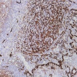
Description and applications: D2-40 recognizes the M2A antigen encoded by the gene Prox1 localised on the chromosomal region 1q32.2–q32.3. It consists in a sialoglycoprotein of 40 kDa, containing mucin-type carbohydrate structure required to preserve the antigenicity of this molecule. M2A is expressed in lymphatic vessels and is absent in venous and arterial vascular endothelium. Other normal tissue that express Podoplanin are myoepithelial cell, mesothelial cells, dendritic cells, cells of the biliary ducts, osteocytes, chondrocytes, granulosa cells, squamous epithelium basal cells etc. It is expressed in various types of vascular tumors, seminomas and in some degree embryonal carcinomas, mesotheliomas, haemangioblastomas, schwanomas, dermatofibromas, thricoblastomas etc.
Composition: anti-Podoplanin mouse monoclonal antibody obtained from supernatant culture and prediluted in a tris buffered solution pH 7.4 containing 0.375mM sodium azide solution as bacteriostatic and bactericidal.
-
 آنتی بادیهای ایمونوهیستوشیمی
آنتی بادی Cyclin E1 (EP126)
Name: Rabbit anti-human Cyclin E1 Monoclonal Antibody clone EP126
Description and applications: This antibody reacts with human protein cyclin E. E-type cyclins (cyclin E1 and cyclin E2) are expressed during the late G1 phase of the cell cycle and until the end of the S phase. Cyclin E activity is limited by the passage of the cells through the “R” restriction point, which marks a “point of no return” for cells going into division from a quiescent state (G0) or through G1 and S phases. Cyclin E expression is mainly regulated by gene transcription by members of the E2F transcription factor family and by degradation through the proteasome catabolic pathway. Cyclin E binds and activates Cdk2 kinase and, through phosphorylation of this kinase’s substrate, the socalled pocket proteins, the cyclin/Cdk2 complex regulates the entry into the S phase by initiating a cascade of events that allows the specific expression of S-phase genes. In addition, cyclin E plays a direct role in the initiation of DNA replication and in the control of genomic and centrosome stability. Overexpression of cyclin E shortens the G1 phase of the cell cycle, accelerating progression to the S phase. High levels of cyclin E expression have been associated with the initiation and progression of different human neoplasms, particularly breast cancer, leukaemias, and lymphomas, among others.
Composition: Anti-human Cyclin E1 rabbit monoclonal antibody purified from serum and prepared in 10mM PBS, pH 7.4, with 0.2% BSA and 0.09% sodium azide
Intended use: Immunohistochemistry (IHC) on paraffin embedded tissues. Not tested on frozen tissues or Western-Blotting
Immunogen: Recombinant protein corresponding to residues from human protein cyclin E.
-
 آنتی بادیهای ایمونوهیستوشیمی
آنتیبادی PARP-1 – پلیکلونال
Name: Poly (ADP-Ribose) Polymerase 1 Antibody
Description and applications: PARP (EC 2.4.2.30) is a nuclear protein with a molecular mass of 116 kDa and two zinc finger motifs which binds to DNA and specifically detects DNA nicks and breaks produced by different genotoxic agents. PARP catalises the ADP-ribosylation of proteins using NAD+ as a substrate. PARP activation is a consequence of ischemic injury and leads to intracellular depletion of NAD+, which can only be replaced by ATP consumption. Ischemia/Reperfusion (I/R) injury results in substantial DNA damage and cells need to consume large amounts of ATP to sustain reparative poly-ADP-ribosylation. Proteolysis of PARP to its stable 85-kDa fragment is an early marker of programmed cell death (apoptosis), mediated by caspase 3 or CPP32. Cleavage occurs between Adp216 and Gly217, a site in PARP conserved across species. This antibody reacts with human, mouse and rat PARP. PARP-1 determination may be useful for detecting cells undergoing DNA replication or repair. In ischemia/reperfusion situations, it may serve as an early marker of cell death. Studies on chemical carcinogenesis have involved PARP on its pathogenesis. Recently, PARP has been considered a therapeutic target in some types of malignancies such as breast carcinomas with BRCA1/2 mutation.
Composition: anti-human Poly (ADP-Ribose) Polymerase 1 rabbit polyclonal antibody purified from serum and prepared in 10mM PBS, pH 7.4, with 0.2% BSA and 0.09% sodium azide
Intended use: Immunohistochemistry (IHC) on paraffin embedded tissues. Not tested on frozen tissues or Western-Blotting
Immunogen: Synthetic peptide derived from the Nterminal region of human protein PARP.
-
 آنتی بادیهای ایمونوهیستوشیمی
آنتی بادی Cytokeratin (CAM 5.2)
Name: Keratin CAM5.2 Antibody Clone CAM5.2
Description and applications: The nomenclature coined in 1982 by Moll and Franke assigned ranges from 1 to 8 for keratin type II (neutral or basic) and between 9 and 21 for type I (acidic). However and because of the high homology among the different molecules is common for a single monoclonal antibody to react with different types of keratins. Keratin CAM 5.2 antibody reacts with 38-50 kD molecular weight keratins present in most simple and ductal epithelia. This antibody is useful for identifying normal and neoplastic epithelial cells in histological sections. Positive stain is obtained in adenocarcinomas of the breast, bowel, liver, renal, neuroendocrine carcinomas (eg Merkel cell tumor), mesotheliomas , germ cell tumors (except seminoma), synovial and epithelioid sarcoma. Some tissues of non-epithelial origin can be immunostained as smooth muscle, some breast sarcomas, meningiomas and malignant neuroblastomas. CAM 5.2 doesn´t stain the stratified epitheliums and does not react with carcinomas arising in the esophagus, vagina and cervix. Negative results were also obtained in lymphomas, melanomas, meningiomas, sarcomas and seminomas.
Composition: IgG2a immunoglobulin, obtained from mouse ascites purified by chromatography and diluted in Tris Buffer, pH 7.3-7.7, with 1% BSA and <0.1% Sodium Azide.
Imunogen: human colorectal carcinoma cell line HT24.
-
 آنتی بادیهای ایمونوهیستوشیمی
آنتی بادی Progesterone Receptor (16)
Name: Mouse Anti-Progesterone Receptor (PR) Monoclonal Antibody clone 16
Description and applications: This mouse monoclonal antibody clone 16 it has been demonstrated to react with both A and B forms of human progesterone receptor by Western blotting procedure. In normal conditions, the nuclear expression of PR occurs in the acinary cells of the breast, endometrium (proliferative phase glands and stroma are strongly positive while absence or variable expression is noted in the secretory phase glands and stroma) myometrium and superficial cervical stroma. PR along with estrogen receptors, Cerb-B2, Ki-67 and p53 are considered to have prognostic value in breast cancer. As the neoplastic growth is hormone dependent, the increased expression of PR is associated with a better response to Tamoxifen-treatment in breast cancers. PR determination is also useful in gynecologic pathology where is positive in endometrial adenocarcinoma, stromal sarcomas or leiomyomas. Meningiomas also show positivity for the PR.
Composition: anti-PR mouse monoclonal antibody obtained from supernatant culture and prediluted in a tris buffered solution pH 7.4 containing 0.375mM sodium azide solution as bacteriostatic and bactericidal.
Immunogen: Recombinant protein encoding N-terminal region of human progesterone receptor.
برای تماس کلیک کنید
سما تشخیص آریا ، عرضه کننده متنوع ترین و کامل ترین محصولات کیت های آزمایشگاهی