-
 ایمونو هیستوشیمی
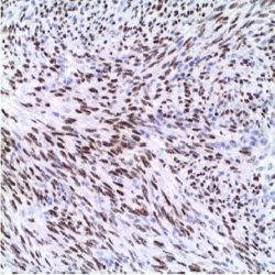
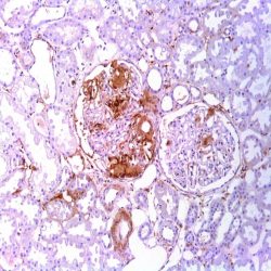
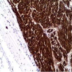
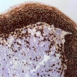
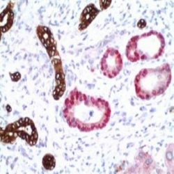
آنتی بادی Human Herpes Virus 8 (13B10)
Name: Herpes Virus 8 (HHV-8) Antibody (Clone 13B10)
Description and applications: Human herpesvirus type 8 (HHV-8) is the likely etiological agent of Kaposi’s sarcoma (KS). HHV-8 DNA sequences have been found in Kaposi’s sarcoma lesions, primary effusion lymphoma, and multicentric Castleman’s disease via polymerase chain reaction and in situ hybridization. Latent nuclear antigen (LNA-1, LNA, LANA-1), also known as ORF73, is a 222- or 234 kD protein that is consistently expressed in HHV-8 infected cells. Anti- HHV-8 labels the latent nuclear antigen protein via immunohistochemistry.
Composition: anti-HHV-8 mouse monoclonal antibody obtained from supernatant culture and prediluted in a tris buffered solution pH 7.4 containing 0.375mM sodium azide solution as bacteriostatic and bactericidal.
-
 آنتی بادیهای ایمونوهیستوشیمی
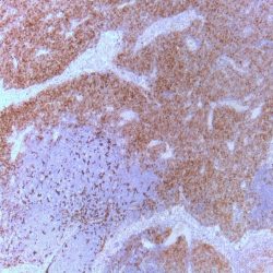
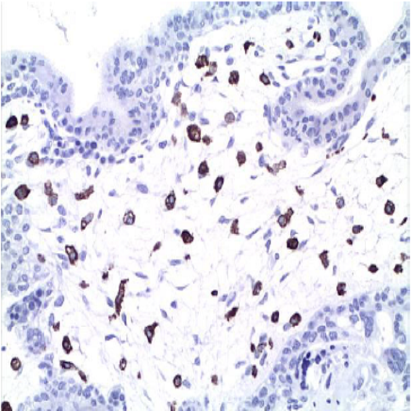
آنتی بادی INI1 (BAF47/SNF5) (25/BAF47)
Name: INI-1 (BAF47/SNF5) Antibody (Clone 25/BAF47)
Description and applications: SWI/SNF complexes facilitate gene activation and transcription factor binding by altering repressive chromatin structures in an ATP-dependent manner. In mammals, SWI/SNF complexes are present in mutliple forms that include 9-12 BRG1-associated factors (BAFs) per complex. These BAF proteins range in molecular weight from 47 to 250 kDa. BAF47/SNF5 (SMARCB1/Ini1) complexes with BRG1- and BRM-containing SWI/SNF complexes. BAF47/SNF5 mRNA is widely expressed in cell lines and tissues, and BAF47/SNF5 gene is mutated in many human tumors. The latter is indicative of a tumor suppressor role for BAF47/SNF5. In addition, mice deficient for BAF47 die early in embryogenesis, while BAF47/SNF5 heterozygous mice display a variety of tumors in the soft tissues of the head and neck. BAF47/SNF5 also binds the HIV-1 integrase and stimulates integrase-mediated DNA joining activity. Thus, BAF47/SNF5 is a component of SWI/SNF complexes that may be critical for normal development and tumor suppression, but may also be a protein utilized for viral DNA integration into host DNA. Expression in normal tissues is ubiquitous. In tumor tissue loss of protein expression SMARCB1 / INI1 gene was first observed in all malignant rhabdoid tumors of childhood of different location (soft tissue, kidney and CNS) and in medullary carcinomas of the kidney. Also loss of expression is observed in 50% of synovial sarcomas, some myxoid chondrosarcoma and myoepithelial carcinomas, almost all epithelioid sarcoma (proximal and conventional variants) and 50% of malignant tumors of peripheral nerves sheat, all presenting rhabdoid morphology. Loss of immunohistochemistry expression is also observed in cases of poorly differentiated chordoma. The antibody is useful in differentiating rhabdoid tumors of other tumors with similar morphology as desmoplastic round cell tumor, rhabdomyosarcoma and Wilms tumor do not show loss of expression of INI1. The antibody also is useful in confirming the diagnosis of medullary carcinoma and discard possible renal pelvis urothelial carcinoma or other carcinomas of the kidney showing expression for INI1. Maintained staining in most carcinomas, epithelioid angiosarcoma, mesotheliomas, melanomas and other tumors or lesions reagents (granulomas) of epithelioid morphology make this useful antibody in the diagnosis of epithelioid sarcoma.
Composition: anti-human INI-1 mouse monoclonal antibody purified from serum and prepared in 10mM PBS, pH 7.4, with 0.2% BSA and 0.09% sodium azide.
Immunogen: mouse BAF47 aminoacids 257-359.
-
 آنتی بادیهای ایمونوهیستوشیمی
آنتی بادی Actin, Muscle Specific (HHF35)
Name: Muscle Specific Actin,Antibody (Clone HHF35)
Description and applications: Actin is a major component of the cytoskeleton. This antibody recognizes actin of skeletal, cardiac, and smooth muscle cells. It is not reactive with other mesenchymal cells except for myoepithelium. Actin can be resolved on the basis of its isoelectric points into three distinctive components: alpha, beta and gamma in order of increasing isoelectric point. Anti- Muscle-Specific Actin recognizes alpha and gamma isotypes of all muscle groups. Non-muscle cells such as vascular endothelial cells and connective tissues are non-reactive. Also, neoplastic cells of non-musclederived tissue such as carcinomas, melanomas, and lymphomas are negative. This antibody is useful in the identification of rhabdoid cellular elements.
Composition: anti-human Muscle Specific Actin mouse monoclonal antibody purified from serum and prepared in 10mM PBS, pH 7.4, with 0.2% BSA and 0.09% sodium azide.
Immunogen: SDS extract of human myocardium
-
 آنتی بادیهای ایمونوهیستوشیمی
آنتی بادی Adipophilin (Polyclonal)
Name: Adipophilin Antibody (Polyclonal)
Description and applications: Sebaceous carcinoma is a relatively uncommon cutaneous malignancy and mimics other malignant neoplasms, such as basal and squamous cell carcinomas, and benign processes, such as chalazions and blepharitis, sometimes resulting in delayed diagnosis and suboptimal treatment. Adipophilin is present in milk fat globule membranes and on the surface of lipid droplets in various normal cell types. Recently, it has been reported that adipophilin was expressed in sebaceous adenomas with a specific pattern: membranous with strong uptake at the periphery of intracytoplasmic lipid vacuoles. Sebaceous carcinomas are also labeled with a similar pattern. Additionally, in cases of poorly differentiated sebaceous carcinoma, adipophilin highlights the sebocytes with a strong membranous labeling of intracytoplasmic lipid droplets. Moreover, xanthelasmas, xanthogranulomas, xanthomas, metastatic renal cell carcinomas were also weakly-to- moderately positive for adipophilin in one study. Expression of adipophilin with a membranous pattern of staining was not seen in any of the other clear cell lesions of the skin, including basal and squamous cell
carcinomas, trichilemmomas, clear cell hidradenomas,or balloon cell nevi. Interestingly, a nonspecific granular uptake of anti-adipophilin was seen in adjacent macrophages, keratohyalin granules of epithelial squamous cells, and some tumor cells.Therefore, this anti-adipophilin is suitable for immunostaining formalin-fixed, paraffin-embedded tissue and is helpful in the identification of intracytoplasmic lipids, as seen in sebaceous lesions. Acinary cell carcinomas of the breast, some liposarcomas, or carcinomas of the colon, lung, pancreas or prostate and strangely the majority of Burkitt lymphomas might also be positive.
Composition:Anti-human Adipophilin rabbit polyclonal antibody purified from serum and prepared in 10mM PBS, pH 7.4, with 0.2% BSA and 0.09% sodium azide.
-
 آنتی بادیهای ایمونوهیستوشیمی
آنتی بادی Amyloid P (EP1018Y)
Name: Amyloid P Antibody clone EP1018Y
Description and applications: Serum Amyloid P (SAP) is a non-fibrillar plasma glycoprotein that belongs to the pentraxin family. It is universally found in amyloid deposits and this is probably due to its specific calcium-dependent binding to motifs present on all types of amyloid fibrils. SAP is also found to prevent fibrillar breakdown by enzymes and it is believed that it helps maintains stability of the amyloid deposits. It has been shown that SAP binds monocytes with high avidity, but does not bind to erythrocytes, NK cells, T lymphocytes or B lymphocytes. SAP production can be induced by exposure to IL-1, IL-6 and IFN-beta. The SAP-inducing activity was neutralized by antibodies to each of the recombinant cytokines. Recognition of these type of amyloid has prognostic and therapeutic implications. The increase in SAP secretion has been documented in different pathologies including neoplasms, rheumatoid arthritis
and CNS diseases. The synthesis of SAP is increased in systemic amyloidosis and is a common component in amyloid deposits. SAP has also been identified in arteriosclerotic lesions.Composition: Anti-human Amyloid P rabbit monoclonal antibody purified from serum and prepared in 10mM PBS, pH 7.4, with 0.2% BSA and 0.09% sodium azide.
-
 آنتی بادیهای ایمونوهیستوشیمی
آنتی بادی ARG-1 (EP261)
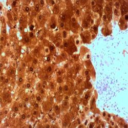
Name: Arginase-1 Antibody (Clone EP261)
Description and applications: Arginase is a manganese metalloenzyme that catalyzes the hydrolysis of arginine to generate ornithine and urea. Arginiase I and II are isoenzymes which differ in subcellular localization, regulation, and possibly function. Arginase I is a cytosolic enzyme, which is expressed mainly in the liver as part of the urea cycle, whereas arginase II is a mitochondrial protein found in a variety of tissues. Antibody to ARG-1 labels hepatocytes in normal tissues and granulocytes in peripheral blood. ARG-1 is a sensitive and specific marker for identification of hepatocellular carcinoma. This antibody is very useful in distinguishing between: 1) liver metastases of various adenocarcinomas and hepatocellular carcinoma (HCC), which can be a realdiagnostic challenge, especially in small biopsies or material from fine needle aspiration ( FNA) and 2) the distinction between different histological variants of HCC and cholangiocarcinoma. Specifically, the ARG-1 antibody is key in the diagnosis of scirrhous hepatocellular carcinoma, where specific markers for adenocarcinomas are generally positive while HepPar-1 in some cases it may be negative. In addition, several studies have shown that ARG-1 antibody is more sensitive than HepPar-1 for immunohistochemical diagnosis of hepatocellular carcinoma while the second is usually negative in poorly differentiated hepatocellular carcinomas and can be positive in adenocarcinomas of pancreatic, gastric, colic origin or even cholangiocarcinoma. However, isolated cases of pancreatic adenocarcinomas and cholangiocarcinomas presented focal staining against ARG-1. In this line, it should be considered that the ARG-1 antibody does not allow differential diagnosis between benign, dysplastic and malignant hepatocyte lesions.
Composition: anti-human ARG-1 rabbit monoclonal antibody purified from serum and prepared in 10mM PBS, pH 7.4, with 0.2% BSA and 0.09% sodium azide.
-
 آنتی بادیهای ایمونوهیستوشیمی
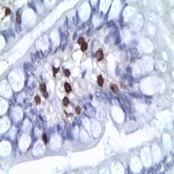
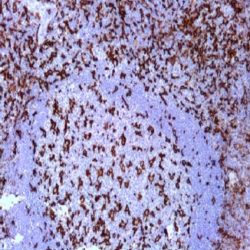
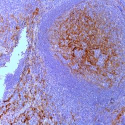
آنتی بادی CD10 (56C6)
Name: CD10 Antibody (Clone 56C6)
Description and aplications: CD10, also known as Common Acute Lymphocytic Leukemia Antigen (CALLA), is a cell surface enzyme with neutral metalloendopeptidase activity which inactivates a variety of biologically active peptides. CD10 is expressed on the cells of lymphoblastic, Burkitt’s, and follicular germinal center lymphomas, and on cells from patients with chronic myelocytic leukemia (CML). It is also expressed on the surface of normal early lymphoid progenitor cells, immature B cells within adult bone marrow and germinal center B cells within lymphoid tissue. CD10 is also present on breast myoepithelial cells, bile canaliculi, fibroblasts, with especially high expression on the brush border of kidney and gut epithelial cells.
Composition: anti-human CD10 mouse monoclonal antibody purified from ascites fluid by Protein A chromatography. Prepared in 10mM PBS, pH 7.4, with 0.2% BSA and 0.09% sodium azide.
-
 آنتی بادیهای ایمونوهیستوشیمی
آنتی بادی CD103 (EP206)
Name: CD103 Antibody (Clone EP206)
Description and applications: CD103, also known as integrin alpha E (ITGAE), is an integrin protein that in humans is encoded by the ITGAE gene. It binds integrin beta 7 to form the complete heterodimeric molecular αEβ7 that binds to an extracellular matrix component and cellular counter receptor. They mediate cell adhesion, migration and signaling and are important for T lymphocyte localization. CD103 is expressed on intraepithelial lymphocytes in mucosal areas, including lung and GI tract. In malignancies, CD103 is present on all enteropathy-type T-cell lymphomas. Additionally, CD103 has been a useful marker for hairy cell leukemia.
Composition: anti-human CD130 rabbit monoclonal antibody purified from ascites. Prepared in 10mM PBS, pH 7.4, with 0.2% BSA and 0.09% sodium azide.
-
 آنتی بادیهای ایمونوهیستوشیمی
آنتی بادی CD117/c-kit (EP10)
Name: CD117/c-kit Antibody (Clone EP10)
Description and applications: This antibody reacts with human oncoprotein c-kit (CD117). The proto-oncogene c-kit encodes a transmembrane receptor with tyrosine kinase activity, c-kit (CD117), which is closely related to the family of platelet derived growth factor receptors. c-KIT is involved in hematopoiesis, gametogenesis and melanogenesis. This antigen is expressed in the normal breast epithelium, melanocytes, mast cells and glia. This antibody is recommended to identify oncoprotein expression c-kit in a variety of normal and neoplastic tissues, including gastrointestinal stromal tumors (GIST). It is also expressed in testicular seminoma, small cell carcinomas of the lung, breast carcinomas, glioblastomas, melanomas, and chronicmyeloid leukemias or acute myeloid leukemias in myeloid blast crisis.
Composition: anti-human CD117/c-kit rabbit monoclonal antibody purified from ascites. Prepared in 10mM PBS, pH 7.4, with 0.2% BSA and 0.09% sodium azide.
-
 آنتی بادیهای ایمونوهیستوشیمی
آنتی بادی CD11b (Integrin Alpha-M) (EP45)
Name: CD11b (Integrin Alpha-M) Antibody (Clone EP45)
Description and applications: Integrin alpha-M (ITAM, ITGAM, CD11b, Mac-1 alpha subunit, C3 alpha chain) is the alpha subunit of the ITAM/beta-2 complex, also named CD11b/CD18 or Mac-1, a leukocyte adhesion heterodimeric glycoprotein. CD11b is involved in monocyte, macrophage, and granulocyte adhesion. CD11b is mainly expressed in myeloid cells of human origin, NK1 cells, monocytes, and granulocytes. In neoplasms, CD11b is useful for the identification of acute myeloid leukaemias (AML) (AML with inv(16) or t(16;16), AML with 11q23/MLL abnormalities, myeloblastic AML with minimal differentiation, myelomonocytic AML, acute monoblastic leukaemia, and acute monocytic leukaemia), where blasts may express CD11b among other molecules of monocytic differentiation. CD11b is also expressed in T-cell large granular lymphocyte leukaemias and in aggressive NKcell leukaemias.
Composition: Anti-human CD11b (Integrin Alpha-M) rabbit monoclonal antibody purified from serum and prepared in 10mM PBS, pH 7.4, with 0.2% BSA and 0.09% sodium azide.
-
 آنتی بادیهای ایمونوهیستوشیمی
آنتی بادی CD11c (Integrin Alpha-X) (EP157)
Name: CD11c (Integrin Alpha-X) Antibody (Clone EP157)
Description and applications: CD11c (ITGAX), a member of the leukointegrin family, shares the same beta subunit with other members of the leukocyte adhesion molecule family, which includes CD11a (LFA-1), CD11b (MAC-1) and CD11d (ITGAD), but has a unique alpha chain. CD11c has been shown to play a role in phagocytosis, cell migration, and cytokine production by monocytes/macrophages as well as induction of T cell proliferation by Langerhans cells. CD11c is expressed prominently on the plasma membranes of monocytes, tissue macrophages, NK cells, and most dendritic cells (DCs). A lower level of expression is also observed on neutrophils as a result of its high level of expression on most DCs. An antibody to CD11c may aid in identification of lesions with histiocytic origin. It may also been used as a marker for hairy cell leukaemia in paraffin embedded tissues.
Composition: anti-human CD11c rabbit monoclonal antibody purified from serum and prepared in 10mM PBS, pH 7.4, with 0.2% BSA and 0.09% sodium azide.
-
 آنتی بادیهای ایمونوهیستوشیمی
آنتی بادی CD13 (EP117)
Name: Rabbit anti-human CD13 Monoclonal Antibody
Composition: anti-human CD13 rabbit monoclonal antibody purified from ascites fluid by chromatography. Prepared in 10mM PBS, pH 7.4, with 0.2% BSA and 0.09% sodium azide
Intended use: Immunohistochemistry (IHC) on paraffin embedded tissues. Not tested on frozen tissues or Western-Blotting
Immunogen: A synthetic peptide corresponding to residues in human CD13 protein
Visualization: Cell membrane
-
 آنتی بادیهای ایمونوهیستوشیمی
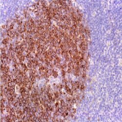
آنتی بادی CD138 (EP201)
Name:CD138 Antibody clone EP201
Description and applications: CD138, also known as Syndecan-1, is a member of the transmembrane heparan sulfate proteoglycan family, acts as an extracellular matrix receptor and is involved in many cellular functions, including cell-cell adhesion and cell-matrix adhesion. CD138 expression is found in both hematopoietic and non-hematopoietic cells. In the hematopoietic system, CD138 labels plasma cells. It is an excellent marker for plasmacytic differentiation within the spectrum of hematologic malignancy. It´s a very usefull tool in the diagnostic of chronic endometritis associated with infertility. Among nonhematolymphoid cells, CD138 reactivity is observed inmany types of epithelial cells and stoma cells in both normal and tumor tissues. These staining should
be remembered and not interpreted as unspecific.Composition:Anti-human CD138 rabbit monoclonal antibody purified from serum and prepared in 10mM PBS, pH
7.4, with 0.2% BSA and 0.09% sodium azideIntended use: Immunohistochemistry (IHC) on paraffin embedded tissues. Not tested on frozen tissues or Western-Blotting
-
 آنتی بادیهای ایمونوهیستوشیمی
آنتی بادی CD14 (EP128)
Name: CD14 Antibody (Clone EP128)
Description and applications:CD14 is a 55-kDa protein found as a glycosylphosphatidylinositol (GPI)- anchored protein on the surface of monocytes, macrophages, and polymorphonuclear leukocytes, and as a soluble protein in the blood. Its main function is to serve as a receptor for lipopolysaccharide (LPS). Besides its role in endotoxin signaling, it has been proposed that CD14 is involved in the transportation of other lipids, cell-cell interactions during different immune responses, and recognition of apoptotic cells.
CD14 is highly expressed on the surface of monocytes/macrophages and strongly up-regulated during the differentiation of monocytic precursor cells into mature monocytes. Therefore, CD14 has been commonly used as a differentiation marker for monocytes/macrophages. An antibody to CD14 also labels Langerhans cells and dendritic cells.Composition: Anti-human CD14 rabbit monoclonal antibody purified from serum and prepared in 10mM PBS, pH 7.4, with 0.2% BSA and 0.09% sodium azide.
-
 آنتی بادیهای ایمونوهیستوشیمی
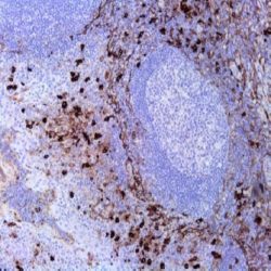
آنتی بادی CD15 (MMA)
Name: CD15 Antibody (Clone MMA)
Description and aplications: This antibody reacts against the human antigen CD15 (fucosyltransferase 4) also known as Lewis X (Lex); 3-fucosyl-Nacetyllactosamine [3-FL]; X hapten; specific embryonic antigen-1 to step [SSEA-1]; lacto-N-fucopentaose III
[LNFP III]. This antibody is expressed in normal myelomonocytic cells (90% of human neutrophil granulocytes in peripheral blood and lymphoid and 30% – 60% of circulating monocytes), not being present in normal lymphocytes. CD15 is consistently expressed by the
epithelial cells of renal tubules, gastric glandular epithelium, tonsil and esophageal squamous epithelium, ductal epithelium of salivary glands, pancreas and mammary ducts, thymus Hassall corpuscles, gray and white matter and in endocrine cells of the adrenal cortex and anterior pituitary. In neoplasms, CD15 antigen is expressed by the Reed- Sternberg cells of classic Hodgkin lymphoma (membrane pattern and / or Golgi area), in some leukemias (acute and chronic myeloid leukemia (> 50%), acute myelogenous leukemia (> 95% of chronic phase and> 50% blast phase), and in carcinomas derived from organs whose cells are positive for CD15, including adenocarcinomas of kidney, digestive tract, squamous cell carcinomas. The CD15 may be useful in confirming the diagnosis of Hodgkin’s disease and the differential diagnosis of mesothelioma (usually negative) and lung adenocarcinoma (usually positive).Composition: anti-human CD15 mouse monoclonal antibody purified from ascites fluid by Protein A chromatography. Prepared in 10mM PBS, pH 7.4, with 0.2% BSA and 0.09% sodium azide.
-
 آنتی بادیهای ایمونوهیستوشیمی
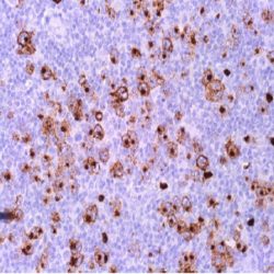
آنتی بادی CD163 (EP324)
Name: CD163 Antibody (Clone EP324)
Description and aplications: CD163 was recently identified as an acute phase-regulated transmembrane protein whose function is to mediate the endocytosis of haptoglobin-hemoglobin complexes. This receptor is expressed on the surface of monocytes (low expression) and tissue macrophages [also known as histiocytes] (high expression). It is a member of the cysteine-rich scavenger receptor superfamily, encoded by a gene localized on human chromosome 12p13.3. Solubilized in plasma, CD163 functions as an anti-inflammatory signal and has many roles in disease processes that range from autoimmune conditions such as rheumatoid arthritis to atherosclerosis. Previous work has shown that the CD163 gene can be regulated by glucocorticoids, IL-10, and other inflammatory modulators, and is highly expressed in inflamed tissues, consistent with its role in the resolution of inflammation.
Staining for CD163 has been helpful in distinguishing synovial macrophages from synovial intimal fibroblasts in the setting of rheumatoid arthritis,where its specificity for macrophages was found to be superior to that of CD68, which does not discriminate between these cell types. Increased levels of CD163 were also detected in patients with microbial infections and myelomonocytic leukemias by an enzyme-linked immunosorbent assay. Flow cytometry studies have confirmed that CD163 expression is limited to leukemias with monocytic differentiation. Another recent study showed that all 5 cases of synovial-type giant cell tumors of the vertebral column stained for CD163.Composition: anti-human CD163 mouse monoclonal antibody obtained from supernatant. Prepared in 10mM PBS, pH 7.4, with 0.2% BSA and 0.09% sodium azide.
-
 Dual Staining
MASTER DUAL STAINING KIT
Available Size : 100 test
Intended Use : Diagnostic in vitro in humans
Description: Master Dual Staining Kit contains reagents to perform manual or automated immunohistoquemical dual
staining on human tissue sections, fixed in buffered formalin and embedded in paraffin.
This kit contains a visualization system based on micropolymers and is sufficient to perform 100 determinations
following the recommended protocol.Limitations Of The Reactives
The use on frozen tissue has not been evaluated.
In order to use the Master Dual Staining kit it should be noted that the two antibodies must come from different
animals (mouse / rabbit).
Due to the presence of a soluble chromogen in alcoholic solutions, the slides must be air-dried and for final mounting
an aqueous mounting media should be used. -
 AP
APMASTER POLYMER PLUS ALKALINE PHOSPHATASE DETECTION SYSTEM
Available Size : 100 test / 500 test
Kit components:
DAB Substrate Buffer (READY-TO-USE)
DAB Chromogen Concentrate (READY-TO-USE)
DAB enhancer (READY-TO-USE)Master Polymer Plus AP is the result of the experience of a decade in systems of polymers for
immunohistochemistry. The detection system has proven greater sensitivity with an increase in the antigenantibody
binding signal. The new technology of micropolymers provides greater advantages as compared to
the conventional immunohistochemistry techniques and facilitates the detection of antigens in the different
cellular compartments (nucleus, cytoplasm, cytoplasmic membrane) due to the smaller size of the molecule.
The technical procedure used eliminates the background staining originated by the non-specific binding to
endogenous biotin molecules, as Master Polymer Plus AP is a procedure not based in the reaction
avidin/biotin.
The detection system Master Polymer Plus AP is highly sensitive, provides a low background staining and
the results obtained are higher than the ones obtained with the conventional procedures of
Streptavidin/Biotin or long polymers.
The system Master Polymer Plus AP is elaborated with the technology of micropolymers.
This system can be used with:
– Monoclonal primary antibodies obtained in mouse.
– Monoclonal and polyclonal primary antibodies obtained in rabbit. -
 DAB
DABImmunoperoxidase DAB Kit (Dark Brown)
Available Size : 100 test / 500 test
Kit components:
DAB Substrate Buffer (READY-TO-USE)
DAB Chromogen Concentrate (READY-TO-USE)
DAB enhancer (READY-TO-USE)Spcificity, Interferences and Limitations:
The Immunoperoxidase DAB Kit (Dark Brown) is preferred for immunohistochemical staining methods, based
on peroxidase marked antibodies and in situ hybridization.
Once the DAB oxidation has occurred, a brown insoluble precipitate is obtained. An increase or decrease in the
ratio between the chromogen and its substrate volumes, as well as exposure to bright light, can affect the
degree of specific staining and background of the final reaction. For this reason it is recommended that the
staining should be made in low light conditions. -
 DAB
DABMaster Polymer Plus Detection System (Peroxidase)
Available Size : 100 test / 500 test / 1250 test
Intended use: Product for in vitro diagnostic use.
Master Polymer Plus Detection System is developed based on the micro-polymer technology and is the result of a decade of experience in polymer systems for immunohistochemistry. This detection system has demonstrated higher sensitivity with an increase in the antigen-antibody binding signal. The novel technology of micro-polymers provides greater advantages than conventional immunohistochemical techniques and facilitates the detection of antigens in different cellular compartments (nucleus, cytoplasm and cell membrane) due to the smaller size of the molecule. It also eliminates background staining caused by nonspecific binding of endogenous biotin molecules as the Master Polymer Plus Detection System staining procedure is not based on the avidin / biotin reaction.
– Primary monoclonal and polyclonal antibodies obtained in mouse
– Primary monoclonal and polyclonal antibodies obtained in rabbit.
برای تماس کلیک کنید
سما تشخیص آریا ، عرضه کننده متنوع ترین و کامل ترین محصولات کیت های آزمایشگاهی