برای تماس کلیک کنید

سما تشخیص آریا ، عرضه کننده متنوع ترین و کامل ترین محصولات کیت های آزمایشگاهی

آنتی‌ بادی Phosphohistone H3 (PHH3) (Polyclonal)

Name: Phospho-Histone H3 (PHH3) Antibody

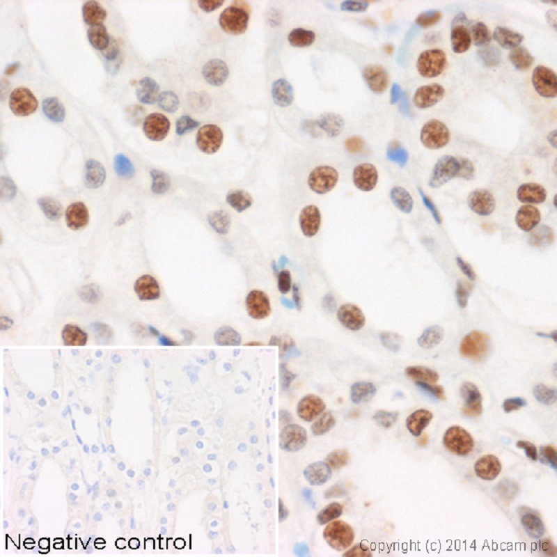
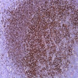
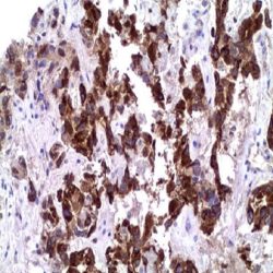
Description and aplications: The phosphohistone H3 (phh3) is the phosphorylated form of nuclear histone H3, which together with the latter one forms the major protein component of the chromatin in eukaryotic cells. In mammalian cells the phosphohistone H3 is negligible during interphase, but reaches its maximum expression at mitosis, more precisely at the transition between the late G2 and M phases of the cell cycle. Immunohistochemical studies have shown that the PHH3 antibody specifically detects the nuclear histone H3 only when protein phosphorylation occurs at the level of serine 10 or serine 28. These studies have also shown that this phosphorylation during apoptosis does not occur on the histone H3, so that the presence of phh3 molecule can serve as a good marker for separating mitotic figures from the apoptotic bodies and nuclear karyorrhexis detritus and is likely to constitute a useful tool for tumor grading, treatment stratification and tumor follow-up especially in CNS, skin, lung, esophagus, breast, soft tissue, stromal gastrointestinal and gynecologic tumors. In fact, recent studies granted PHH3 with greater importance and relevance than Ki- 67 in identifying mitotic figures and a higher intra -and inter reproducibility in cases of melanoma.

Composition: anti- PHH3 rabbit polyclonal antibody obtained from supernatant culture and prediluted in a Tris buffered solution pH 7.4 containing 0.375mM sodium azide solution as bacteriostatic and bactericidal. The quantity of the active antibody was not determined.

Intended use : Immunohistochemistry (IHC) on paraffin embedded tissues. Not tested on frozen tissues or Western-Blotting

حجم موجود: 3 / 7 / 12 میلی‌لیتر

مورد استفاده: ایمونوهیستوشیمی روی بافت پارافین. بر روی بافتهای فروزن یا وسترن بلات تست نشده است.

کلون: Polyclonal

ایزوتایپ: Rabbit IgG

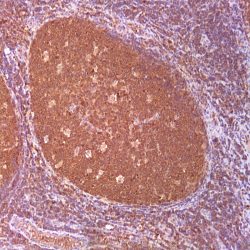
کنترل مثبت: لوزه

قابلیت دیده شدن: هسته

روش پیشنهادی IHC:

  • برش تهیه شده با ضخامت μm4 باید روی لام شارژ شده در طول شب و در دمای 60 درجه سانتی گراد خشک شود.
  • دپارافینه کردن، رهیدراته کردن و استفاده از حرارت جهت بازیابی اپی توپ : بافت را با استفاده از بافر EDTA pH 8 برای بیست دقیقه در دمای 95 درجه سانتی گراد بجوشانید. سپس 5-3 بار با استفاده از آب مقطر یا دیونیزه شده شسته و سپس بیست دقیقه در دمای اتاق خنک کنید.
  • بلاک پروکسیدازهای درونی : بلاک با استفاده از محلول پروکسیداز به مدت ده دقیقه
  • آنتی بادی اولیه: برای 10 دقیقه در انکوبه کنید پروتکل ممکن است بسته به تهیه نمونه و کاربرد خاص متفاوت باشد. شرایط استاندارد باتوجه به آزمایشگاه و کاربر متفاوت می باشد.
  • برای رنگ آمیزی ، می توانید از کیت Master Polymer Plus Detection یا کیتهای دیتکشن سایر کمپانی ها استفاده کنید.
  • رنگ میزی نهایی با هماتوکسیلین و فیکس نهایی اسلاید.

شرایط نگهداری و پایداری : حداکثر 18 ماه؛ در دمای 2-8 درجه سانتیگراد نگهداری شود. فریز نشود.


این آنتی‌بادی متعلق به پنل‌‌‌‌‌‌‌های تشخیصی DERMATOPATHOLOGY و SOFT TISSUE  است.


Download Cataloge

Icon

Phosphohistone H3 (PHH3) (Polyclonal) 344.31 KB 15 downloads

...
.well.c2a3 .btn { padding: 11px 30px; font-size: 11pt; } .well.c2a3 .media-body { font-size: 11pt; } .well.c2a3 .wpdm_icon { height: 42px; width: auto; }

Reviews

There are no reviews yet.

Be the first to review “آنتی‌ بادی Phosphohistone H3 (PHH3) (Polyclonal)”

Your email address will not be published. Required fields are marked *

Scroll to Top